
Tuberous Sclerosis Complex
Metabolic Imaging Biomarkers for TSC
Posted May 9, 2019
Carmen Priolo, M.D., Ph.D., Brigham and Women's Hospital


Tuberous sclerosis complex (TSC) is an autosomal dominant disease characterized by neurologic manifestations and benign tumors of the brain, skin, heart, and kidneys. Up to 80% of women with TSC develop lymphangioleiomyomatosis (LAM), a progressive disease in which muscle-like cells that line the lungs’ airways and blood vessels begin to grow out of control and over time, can destroy the normal lung tissue. Clinical trials showed that the mTORC1 inhibitor rapamycin stabilizes pulmonary function; however, lung function declines and tumor growth resumes when the drug is discontinued. This points to the need to develop and optimize therapeutic strategies in LAM and TSC. A very important component of clinical trial design and implementation is having specific biomarkers of disease progression and response to therapy which currently is not available in the TSC field. Dr. Carmen Priolo’s long-term research goal is to address this need, as well as to understand the biochemical basis of TSC tumorigenesis.
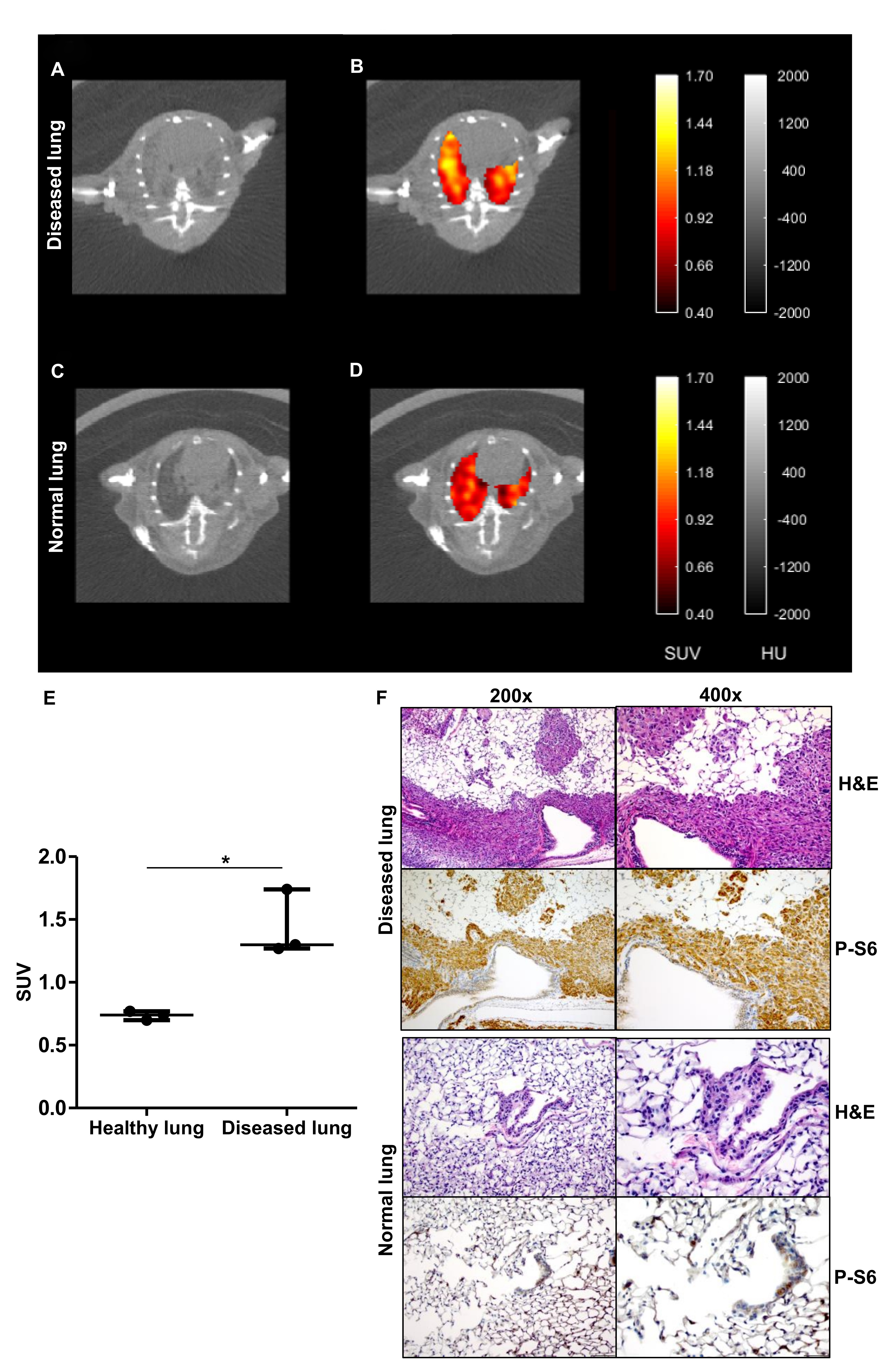
With support from a Fiscal Year 2012 (FY12) and FY15 Tuberous Sclerosis Complex Research Program Exploration - Hypothesis Development Award, Dr. Priolo first sought to answer the questions, “What are the major nutrients used by TSC tumor cells to grow and proliferate? What are the major fuels of TSC tumors? How is utilization of these fuels impacted by rapamycin?” To start, she used metabolic assays to identify nutrients that are preferentially processed by TSC2-deficient cells. She found that some of these nutrients are important for tumor cells to grow and double and are utilized by TSC tumors to make macromolecules or generate energy. Dr. Priolo then teamed up with the Gordon Center for Medical Imaging at the Massachusetts General Hospital, Boston, to chemically-modify specific nutrients (choline and acetate) to generate radioactively labeled derivatives [18F]fluorocholine (FCH) and [18F]fluoroacetate (FACE) to test their potential as biomarkers. Positron Emission Tomography (PET) imaging uses radiolabeled derivatives of nutrients to noninvasively detect (image) body cells that have an increased uptake and consumption of these nutrients. Using PET imaging in preclinical models of TSC and LAM, Dr. Priolo and her team showed that TSC2-deficient tumors exhibited rapid uptake of [18F]FCH and [18F]FACE. These findings provide evidence for testing the potential of [18F]FCH and [18F]FACE as metabolic imaging biomarkers in TSC and LAM.
Referring to what has contributed to her success, Dr. Priolo has stated, “I believe that our strength resides in the multidisciplinarity of our team, which includes physician-scientists, molecular biologists, physicists, and chemists. We brainstormed together to find solutions and set up a platform to test TSC tumor metabolic features generating consistent and rigorous preclinical data that may serve as the first required step to move as quickly as possible to clinical studies. We will be able to use this platform for future studies not only to identify the best potential metabolic biomarkers of TSC, but also to increase our understanding of the molecular mechanisms involved in TSC tumorigenesis.”
Going forward, Dr. Priolo plans to design clinical trials to test the imaging methodologies she has developed in TSC. Some of the tracers she has tested have already been approved by the FDA for the diagnosis of other diseases, including prostate cancer and heart diseases, while other tracers will require special approvals to be brought to the clinics. However, all of these tracers have been shown to be safe for humans through clinical research studies.
In collaboration with the Gordon Center for Medical Imaging, and with support from a LAM Patient Benefit Grant (LAM Foundation), Dr. Priolo is moving from the preclinical to the clinical phase. Dr. Priolo notes that: “Funding from the DoD TSCRP (FY12 and FY15) have been truly critical to support our work.”

Publications:
(1) Verwer EE, Kavanagh TR, Mischler WJ, Feng Y, Takahashi K, Wang S, Shoup TM, Neelamegam R, Yang J, Guehl NJ, Ran C, Massefski W, Cui Y, El-Chemaly S, Sadow PM, Oldham WM, Kijewski MF, El Fakhri G, Normandin MD, and Priolo C. 2018. [18F]Fluorocholine and [18F]fluoroacetate PET as imaging biomarkers to assess phosphatidylcholine and mitochondrial metabolism in preclinical models of TSC and LAM. Clin Cancer Res. 24(23):5925-5938.
(2) Priolo C, Ricoult SJ, Khabibullin D, Khabibullin D., Filippakis H., Yu J., Manning B.D., Clary Clish C., and Elizabeth P. Henske E.P. 2015. Tuberous sclerosis complex 2 loss increases lysophosphatidylcholine synthesis in lymphangioleiomyomatosis. Am J Respir Cell Mol Biol. 53(1):33-41.
Links:
Public and Technical Abstract: Novel Metabolic Biomarkers in Tuberous Sclerosis Complex
Public and Technical Abstract: Novel Application for 18F-Fluorocholine PET Imaging in TSC
Last updated Wednesday, March 12, 2025