
Tuberous Sclerosis Complex
Causes of Epilepsy in TSC- Potential Therapeutic Targets
Posted November 13, 2019
Angelique Bordey, Ph.D., Yale University School of Medicine
Tuberous Sclerosis Complex (TSC) is a multi-organ system disorder; however, the neurological symptoms, specifically epilepsy, cause significant disabilities and have the greatest impact on a patient’s quality of life. Currently, TSC-related epilepsy is treated with either the drug everolimus or invasive brain surgery. Everolimus, an mTORC1 (mechanistic target of rapamycin complex 1) pathway inhibitor, requires life-long treatment and has produced grade 3 or 4 side effects in patients,1 including development of kidney failure, which could exacerbate an existing problem for many TSC patients. Therefore, if cortical tubers are operable most patients choose brain surgery; however, only 50% of TSC patients will be able to properly manage their seizures after removal of cortical tubers due to tuber recurrance.1 Thus, there is an urgent need to understand the mechanism of seizure generation to improve epilepsy treatment options for TSC patients.
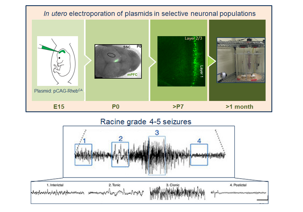
With support from a Fiscal Year 2009 (FY09) Idea Development Award (IDA), Dr. Angelique Bordey and her team from the Yale University School of Medicine generated a novel focal cortical malformation (FCM) mouse model in which Rheb, the activator of mTORC1, is constitutively active (RhebCA), resulting in the formation of cortical tubers similar to those associated with recurrent seizures in TSC patients. Using this model, Dr. Bordey further identified the ectopic expression of a pacemaker channel, HCN4, in tuber neurons, which is not observed in a normal cortex, and postulated that these channels are involved in TSC-related seizures.
With support from an FY15 IDA, Dr. Bordey and her team aimed to expand on their previous research. They sought to evaluate hyperpolarization-activated ion channel isoform 4, HCN4, expression in mouse and human neurons and determine if blocking the activation of the HCN4 channel can serve as a therapeutic target to eliminate TSC-related epilepsy. Using video-EEG recordings in 3- to 5-month-old mice containing FCM, Dr. Bordey confirmed hyper-excitability and seizure activity in the FCM mouse model. Furthermore, patch clamp recordings of acute brain slices in RhebCA neurons showed depolarized resting membrane potentials, which were normalized in the presence of HCN channel blocker, zatebradine, thus validating Dr. Bordey’s hypothesis that HCN4 channels play a role in neuron hyperexcitability. Immunostaining of mouse RhebCA neurons and cortical tissue from a TSC patient that underwent brain surgery as a result of cortical tubers revealed abnormal HCN4 expression in both mouse and human neurons. To evaluate whether blocking HCN4 activation prevents seizure activity in a TSC model, Dr. Bordey introduced nonfunctional HCN4 channels into RhebCA neurons that block the activity of endogenous HCN4 channels. Patch clamp recordings of RhebCA neurons expressing nonfunctioning HCN4 showed a reduction in HCN current, indicating successful blocking and inactivation of these channels. Furthermore, video-EEG recordings of these mice revealed that nonfunctional HCN4, together with RhebCA, expression eliminated seizure activity and confirmed the potential therapeutic benefit of targeting HCN4 channels using gene therapy.
Epilepsy and neurological disabilities are the most debilitating manifestations of TSC and drastically impact a patient’s quality of life. Dr. Bordey’s work provides hope that a new therapeutic target to reduce recurring seizures in TSC patients now exists. Ultimately, targeting HCN4 channels could reduce the number of invasive brain surgeries TSC patients have to undergo.
Reference:
1 Bollo RJ, Kalhorn SP, Carlson C, Haegeli V, Devinsky O, and Weiner HL. 2008. Epilepsy surgery and tuberous sclerosis complex: Special considerations. Neurosurg. Focus. 25, E13.
Publications:
Hsieh L, Wen J, Nguyen L, Torres-Reveron J, Spencer D, and Bordey A. Ectopic HCN4 expression drives mTOR-dependent epilepsy. Manuscript in preparation.
Zhang L, Huang T, Teaw S, and Bordey A. 2019. Hypervascularization in mTOR-dependent focal and global cortical malformations displays differential rapamycin sensitivity. Epilepsia. 60:1255-1265.
Heish L, Wen J, Claycomb K, Huang Y, Harrsch F, Naegele JR, Hyder F, Buchanan G, and Bordey A. 2016. Convulsive seizures from experimental focal cortical dysplasia occur independently of cell misplacement. Nature Communications. 1:7:11753.
Links:
Public and Technical Abstracts: Epilepsy Causes and Treatment in TSC
Last updated Friday, March 7, 2025