
Posted January 14, 2014
Angelique Bordey, Ph.D., Yale University, New Haven, Connecticut
 Tuberous sclerosis complex (TSC) is a genetic disorder resulting from mutations in TSC1 or TSC2 genes. TSC patients develop lesions on multiple organs including the brain, which account for the neurological symptoms of the disease including seizures, mental retardation, and autism. Currently, there is no cure for TSC, and the mechanisms leading to TSC brain lesions and the subsequent development of neurological symptoms are not well defined. Moreover, transgenic animal models of TSC so far have not fully replicated the human pathology.
Dr. Angelique Bordey, through funding from a Fiscal Year 2009 Idea Development Award, developed a unique mouse model of TSC in which Tsc1 is selectively knocked out in developing neurons; this model mimics the human pathology and allows researchers to study the etiology of TSC lesion formation. Dr. Bordey demonstrated that the knockout of Tsc1 in select groups of neurons in embryonic mice already carrying a mutant Tsc1 allele (Tsc1flox/mutant) led to the development of cortical tuber-like lesions that share many characteristics with the cortical lesions seen in the human disease (see Figure 1). Further, her team has demonstrated that Tsc1 deletion in neonatal cells in the subventricular zone results in lesions in the olfactory bulb containing ectopic Tsc1null neurons with abnormal morphology and synaptic connectivity.
Using this mouse model, Dr. Bordey and her research team have identified several proteins, such as hypoxia inducible factor 1 (Hif1a), which are abnormally regulated in TSC. Briefly, they demonstrated that Hif1a plays a role in the survival of newborn diseased neurons in olfactory bulb lesions. Also, when Hif1a levels are increased, there is a significant increase in the basal dendrite complexity and length, a defect that is seen in TSC neurons and contributes to lesion formation. These results suggest that Hif1a may play an important role in the formation of lesions in TSC. Dr. Bordey believes the identification of TSC-related molecules is an important breakthrough that will move the field of developmental biology and TSC forward. Future research by Dr. Bordey and her team will attempt to identify additional proteins that are abnormally regulated in TSC and also determine whether or not restoring Hif1a or other molecules to normal levels can prevent the formation of lesions in TSC.
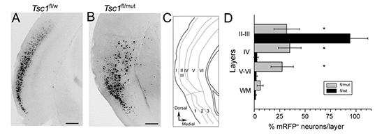
Figure 1. Single-cell Tsc1 deletion at embryonic day 15 generates tuber-like lesions in Tsc1flox/mutant mice. Photograph of neurons expressing red fluorescent protein (mRFP) in cerebral cortex layers II/III from control Tsc1flox/wt (wt: wild type) (A) and Tsc1flox/mutant (B) mice on post-natal day 28 (P28). (C) Schematic of cerebral cortex layers. (D) Bar graph illustrating the number of mRFP neurons in the various cerebral cortex layers of Tsc1flox/wt (gray) and Tsc1flox/mutant (black) P28mice. In the normal mouse brain, mRFP-expressing cell distribution is primarily localized to layers II/III of the cerebral cortex. Single-cell Tsc1 deletion in Tsc1flox/mutant mice resulted in an abnormal distribution of mRFP-expressing neurons across all layers of the cerebral cortex.
Publications:
Feliciano DM, Su T, Lopez J, Platel JC, and Bordey A. 2011. Single-cell Tsc1 knockout during murine corticogenesis generates tuber-like lesions and reduces seizure threshold without astrogliosis. Journal of Clinical Investigation 121:1596-1607
Feliciano DM, Quon JL, Su T, Taylor MM, Bordey A. 2012. Postnatal neurogenesis generates heterotopias, olfactory micronodules and cortical infiltration following single-cell TSC1 deletion. Human Molecular Genetics 21:799-810.
Feliciano DM, Zhang S, Quon JL, and Bordey A. 2013. Hypoxia-inducible factor 1a is a Tsc1-regulated survival factor in newborn diseased neurons. Human Molecular Genetics 22:1725-1734.
Feliciano DM and Bordey A. 2013. Newborn cortical neurons: only for neonates? Trends in Neuroscience 36:51-61.
Feliciano DM, Hartman NW, Lin TV, Bartley C, Kubera C, Hsieh L, Lafourcade CA, O�Keefe R, and Bordey A. 2013. A circuitry and biochemical basis for tuberous sclerosis symptoms: From epilepsy to neurocognitive deficits. International Journal of Developmental Neuroscience pii: S0736-5748.
Lafourcade CA, Lin TV, Feliciano DM, Zhang L, Hsieh LS, and Bordey A. 2013. Rheb activation in subventricular zone progenitors leads to heterotopias, ectopic neuronal differentiation, and rapamycin-sensitive olfactory micronodules and dendrite hypertrophy of newborn neurons. Journal of Neuroscience 33:2419-31.
Links:
Public and Technical Abstracts: Understanding the Etiology of Tuberous Sclerosis Complex