
Peer Reviewed Orthopaedic
Developing and Using a Large Animal Survival Model to Study Treatment for Intra-Articular Fracture-Induced Posttraumatic Osteoarthritis


Posted April 26, 2017
Jessica E. Goetz, Ph.D., University of Iowa, Iowa City, Iowa; Yuki Tochigi, M.D., Ph.D., University of Iowa, Iowa City, Iowa

Intra-articular fracture (IAF) injury is one of the most frequent causes of post-traumatic osteoarthritis (PTOA). Because the IAF injury involves the articular surface of bones of the joint, the entire joint space, cartilage, and bones are all affected. This can lead to complications including uneven joint surfaces, perpetual joint surface damage and misalignment, and joint instability. It is well known that the biomechanical effects of joint trauma and uneven wear put patients at a high risk for the development of PTOA. While IAF mouse models are currently used by researchers to study the progression and treatment of PTOA, these models do not recreate the magnitude of loading seen in larger animals, and their joints are too small to allow treatment of the fracture. Human IAFs are most often treated surgically, and thus surgical treatment is a critical factor to include in studies of development and treatment of PTOA. Due to the need for a human-equivalent weight and a large joint in which surgical fracture treatment can be performed, a large animal survival model of IAF is needed for injury and treatment studies.
To address this gap, Dr. Yuki Tochigi from the University of Iowa was awarded the Fiscal Year (FY) 2009 Technology Development Award from the Peer Reviewed Orthopaedic Research Program (PRORP). The team proposed to study Yucatan minipigs, which reach weights of 150-200 lbs when mature and which have relatively large joints with anatomy similar to human joints. Dr. Tochigi's team developed a surgical technique simulating injury to the ankle of an anesthetized minipig, creating an intra-articular fracture of the distal tibia. After treating the fracture with surgical open reduction and internal fixation (the standard-of-care monitoring of human IAF patients), the team followed the animals' 12-week recovery regimen. The team noted that the minipigs' fractured limbs healed firmly and were capable of physically carrying weight to pre-surgical levels. However, despite these excellent fracture healing outcomes, cartilage damage, including erosion and discoloration developed. Further analysis showed trends of cytokine elevation in the minipigs were similar to human IAF patients, predicting further cartilage damage and eventual PTOA.
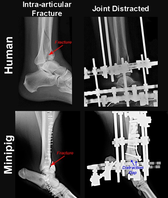
Dr. Jessica Goetz from the University of Iowa resumed the project as the Principal Investigator of the Technology Development Award after Dr. Tochigi transferred to another institution. Results from the project funded by the original award yielded a valuable large animal survival model of IAF, which enabled Dr. Goetz to further study IAF-induced osteoarthritis. In FY 2014, Dr. Goetz received a PRORP Expansion Award to expand on the Technology Development Award for a new project to use her minipig IAF animal model to determine whether joint distraction treatment after IAF could better prevent joint degeneration and development of PTOA. The team designed an external fixator to distract the joint symmetrically by slightly pulling the cartilage surfaces of the minipig's ankle joint apart, thereby increasing the joint space with a goal of protecting the cartilage from load and promoting healing. The result of this procedure is shown in the figure below, in which one can see the space created between the lower leg and foot bones in the minipig ankle that is not present between those bones before the external fixator was applied. The first of two sets of surgeries were completed within the first year of the project, focusing on acute treatment. Results from pending experiments will hopefully show whether timely administered joint distraction treatment could help prevent osteoarthritis progression for IAF patients.
Publications (from work performed under FY09 Technology Development Award):
Tochigi Y, Buckwalter JA, Martin JA, et al. 2011. Distribution and progression of chondrocyte damage in a whole-organ model of human intra-articular fracture. J Bone Joint Surg Am. 93(6):533-539.
Tochigi Y, Zhang P, Rudert MJ, et al. 2013. A novel impaction technique to create experimental articular fractures in large animal joints. Osteoarthritis Cartilage. 21(1):200-208.
Diestelmeier BW, Rudert MJ, Tochigi Y, et al. 2014. An instrumented pendulum system for measuring energy absorption during fracture insult to large animal joints in vivo. J Biomech Eng. 136(6):064502.
Goetz JE, Fredericks D, Petersen E, et al. 2015. A clinically realistic large animal model of intra-articular fracture that progresses to post-traumatic osteoarthritis. Osteoarthritis Cartilage. 23(10):1797-805.
Links:
Last updated Wednesday, March 12, 2025