 An official website of the United States government
 ) or https:// means you've safely connected to the .mil website. Share sensitive information only on official, secure websites.
Peer Reviewed Medical
Promoting Regeneration and Healing During Bone Transport Using a Novel Implant Technology


Posted September 27, 2023
Yunzhi Peter Yang, Ph.D., Stanford University
Dr. Yunzhi Peter Yang
(Photo Provided)
Lower extremity fractures among military personnel caused over 700,000 limited duty days and $116 million in total costs, including $24 million in direct medical expenses, in 2017.1 Fractures, often caused by traumatic injuries, may produce large defects in the bone that the body cannot heal itself, requiring surgical intervention.
Through a process known as bone transport, a surgeon removes the damaged area of bone, and then performs an osteotomy (bone cut) at a point where the bone is healthy and intact. This results in a free segment of bone that can be connected to an external fixation system and moved in a controlled fashion. Over the course of several months to over a year, this segment of bone is slowly moved across the gap created by the damaged bone via adjustments to the external fixation frame device. As the bone segment is pushed across the gap, new bone growth occurs behind the end that was cut. Once the section reaches the far side of the gap, the bone segment fuses to the other length of remaining bone in a process called docking site union. The new bone bridging the gap then hardens over several more months, and the remedy of the defect is complete. However, sometimes the bone segment does not fuse properly; this is known as docking site nonunion, which requires a secondary surgery to repair. In addition, the external fixation device limits functionality, and the pins anchoring the device in the bone are susceptible to infection.
With support from a fiscal year 2021 Peer Reviewed Medical Research Program Technology/Therapeutic Development Award - Funding Level 1, Dr. Yunzhi Peter Yang's team at Stanford University aims to validate a novel implant technology to support better regeneration and faster healing during bone transport procedures; they are currently in year two of their four-year period of performance. The team developed intramedullary (IM) implants, which function as nails to anchor the fixation device to the bone and release bone morphogenetic protein (BMP)-2. Previous studies showed that BMPs, which are bone growth factors that promote healing, accelerate bone regeneration in animal models.2 To improve the protein's effects and delivery, the team designed the IM implants to slowly release BMP-2 into the bone and surrounding tissue, providing a controlled and sustained dose throughout the bone transport process. Internal fixation devices, like IM implants, reduce the use of external frames and ease patient burden. Of note, the IM implants are usually made of metal and may require removal surgery after bone transport is complete.3 To reduce the need for this follow-up surgery, Dr. Yang's team constructed their novel implants from U.S. Food and Drug Administration-approved biodegradable materials.
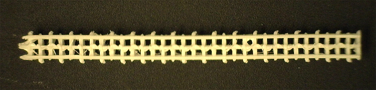 Dr. Yunzhi Peter Yang and his team at Stanford University developed an intramedullary implant device, which anchors to the bone and releases bone morphogenetic protein, found to promote healing and regeneration in the bone. Dr. Yang received a fiscal year 2021 Technology/Therapeutic Development Award - Funding Level 1 from the Peer Reviewed Medical Research Program to research the validation of the technology of this device. (Figure Provided)
As part of the PRMRP Technology/Therapeutic Development Award, the team first tested the BMP-2-releasing, biodegradable implants in a rat model of bone transport to evaluate their effect on bone growth and healing. As compared to standard devices, the implants releasing BMP-2 significantly improved bone volume and tissue volume at the regenerative site and at the docking site, increasing mechanical strength and bone fusion. With confirmation of improved outcomes in the animal model, the team plans to test the implants in a preclinical large animal model of bone transport. Toward this goal, they scaled the size of the implants using 3D-printing, optimizing their physical and mechanical properties. They also evaluated the release of BMP-2 in the new implants, selecting the optimal dose for a large animal study. Once the design and characteristics are finalized, the team will first test conventional techniques and commercially available bone grafting materials during bone transport surgery to repair the metatarsus, or lower leg bone. They will then test the BMP-2-releasing biodegradable implants, measuring healing and mechanical strength of the new bone to determine whether they lead to improved bone transport outcomes.
If preclinical testing is successful, Dr. Yang's team will be well positioned to move this novel technology to clinical trials in future awards. Ultimately, if proven acceptable for clinical use, these implants could lead to improved bone transport procedures and better functional outcomes for those impacted by significant bone injuries.
References:
1Forest L. 2021. The cost of lower extremity fractures among active duty U.S. Army soldiers, 2017. Military Health System. https://pubmed.ncbi.nlm.nih.gov/34379379/.
2Li Y, Lui R, Hu J, et al. 2016. Recombinant human bone morphogenetic protein-2 suspended in fibrin glue enhances bone formation during distraction osteogenesis in rabbits. Archives of Medical Science 12(3):494:501. https://doi.org/10.5114/aoms.2016.59922.
3Prediger B, Mathes T, Probst C, and Pieper D. 2020. Elective removal vs. retaining of hardware after osteosynthesis in asymptomatic patients - a scoping review.
Publications:
Lin S, Maekawa H, Moeinzadeh S, et al. 2023. An osteoinductive and biodegradable intramedullary implant accelerates bone healing and mitigates complications of bone transport in male rats. Nature Communications 14(1):4455. https://doi.org/10.1038/s41467-023-40149-5.
Links:
Last updated Friday, December 19, 2025