 An official website of the United States government
 ) or https:// means you've safely connected to the .mil website. Share sensitive information only on official, secure websites.
Peer Reviewed Medical
Investigating the Cellular and Molecular Mechanisms Associated with Worsened Outcomes in COVID-19 Patients with Chronic Lung Disease


Posted August 10, 2021
Jonas Schupp, M.D., Yale University
Jonathan Kropski, M.D., Vanderbilt University Medical Center
Nicholas Banovich, Ph.D., Translational Genomics Research Institute

Dr. Jonas Schupp
The coronavirus disease 2019 (COVID-19) is caused by severe acute respiratory syndrome coronavirus 2 (SARS-CoV-2) and has emerged as a global pandemic. Chronic lung diseases, such as pulmonary fibrosis (PF), are a significant comorbidity in COVID-19 patients, making them more vulnerable to severe disease and poor outcomes. However, the molecular mechanisms associated with the increased risk are not well understood. PF is a severe lung disease that results in irreversible scarring of the lung tissue that worsens overtime. With their existing awards, two Peer Reviewed Medical Research Program-funded teams pivoted their studies to help answer questions regarding the molecular pathways of the lung microenvironment, particularly in PF and idiopathic pulmonary fibrosis (IPF) lungs, and how they relate to SARS-CoV-2 viral entry and COVID-19 disease susceptibility.
Dr. Jonas Schupp was awarded a Fiscal Year 2018 (FY18) PRMRP Discovery Award to study the cellular and genetic regulatory networks in the microenvironment of the lung to understand IPF disease progression and identify potential biomarkers and drug targets. Under the mentorship of Dr. Naftali Kaminski, Dr. Schupp’s team at Yale University optimized a single nuclei RNA sequencing method to characterize the IPF lung. They also developed an automated computational pipeline to relate cellular transcriptional profiles to immune function and disease progression, which was tested on sputum samples from cystic fibrosis patients, and the results were published in the American Journal of Respiratory Critical Care Medicine. Dr. Schupp’s team will use this new data processing method to analyze differentially affected regions within IPF lungs, which will allow them to understand the cell-type-specific regulatory networks and pathways related to disease pathogenesis and identify biomarkers and targets for novel therapeutics.

Dr. Jonathan Kropski
While investigating the gene expression patterns that drive PF pathogenesis with the support of a FY18 PRMRP Investigator-Initiated Research Award – Partnering PI Option, Drs. Jonathan Kropski and Nicholas Banovich began examining whether gene expression programs associated with entry, infection, and response to SARS-CoV-2 are altered in patients with PF. They collected and sequenced samples from highly fibrotic portions of 20 PF lung samples, and began analyzing single-cell RNA sequencing (scRNAseq) data to identify cell-type-specific gene expression changes associated with PF. The research team will continue profiling the cellular and molecular landscape of PF lungs from at least 75 patients to determine the gene expression patterns that contribute to disease progression.
The PRMRP-supported work by Drs. Kropski, Banovich, and Schupp ultimately led to a comprehensive analysis, led by Dr. Linh Bui at Translational Genomics Research Institute and Dr. Nichelle Winters at Vanderbilt University Medical Center and reported in a recent bioRxiv paper, outlining the association of chronic lung diseases with gene expression profiles that aid viral entry of SARS-CoV-2 and COVID-19 disease severity. The transcriptomes of over 600,000 single cells isolated from healthy control lung samples, as well as lung samples from patients with chronic obstructive pulmonary disease (COPD), IPF, and other interstitial lung diseases, were analyzed to identify molecular characteristics of lung cells that could account for worse COVID-19 outcomes. The investigators found that the SARS-CoV-2 entry receptor angiotensin-converting enzyme 2 (ACE2) and viral entry-associated protease transmembrane protease, serine 2 (TMPRSS2) had similar cellular distribution and relative expression in disease and control lungs, suggesting that changes in the expression of SARS-CoV-2 entry factors do not play a major role in disease severity. However, isolated type 2 alveolar (AT2) cells from disease lungs expressed higher levels of genes directly linked to viral replication processes and innate immune response. In addition, the investigators identified unique ACE2-correlated gene profiles for each disease group in AT2 cells and subsequently measured the levels of ACE2 in various regions of the lung. Notably, ACE2 expression was elevated in IPF small airway sections, which could serve as an indicator for a more severe localized viral response.

Dr. Nicholas Banovich
The research team then examined the immune cell population in the lung to determine whether immune dysregulation in patients with pre-existing chronic lung disease contributes to COVID-19 severity. Disease lungs had a significant increase in the proportion of CD4, CD8, and NK cells when compared to control lungs and many of the immune cells showed upregulation of IL-6 and MHC class II genes. These differences in inflammatory gene expression in disease lungs compared to healthy lungs indicate how chronic lung disease alters the inflammatory microenvironment of the lung after SARS-CoV-2 infection, contributing to increased risk of severe disease. These findings suggest that the immune microenvironment in the lungs of patients with chronic lung disease is dysregulated and may prime the lung epithelium for more severe infection and worse outcomes in COVID-19.
As part of the Human Cell Atlas (HCA) Biological Network, Drs. Kropski and Banovich also contributed to early findings related to SARS-CoV-2 published in Nature Medicine. The paper outlines multiple scRNAseq datasets generated within the HCA consortium and other resources that highlight the potential role of genes involved with the initial SARS-CoV-2infection, spread, and clearance. The investigators found that ACE2 and TMPRSS2 are highly expressed in nasal epithelial cells and are co-expressed with genes involved in innate immunity.
The work by these PRMRP-funded investigators has provided insight into the cellular and molecular mechanisms that make patients with chronic lung disease more susceptible to severe COVID-19 and poorer outcomes. Key findings showed that dysregulation of genes related to viral replication and the innate immune response in lung epithelial cells, as well as differences in gene expression profiles of the inflammatory microenvironment, may be primary factors for understanding the increased risk for hospitalization and mortality in COVID-19 patients with pre-existing lung diseases.
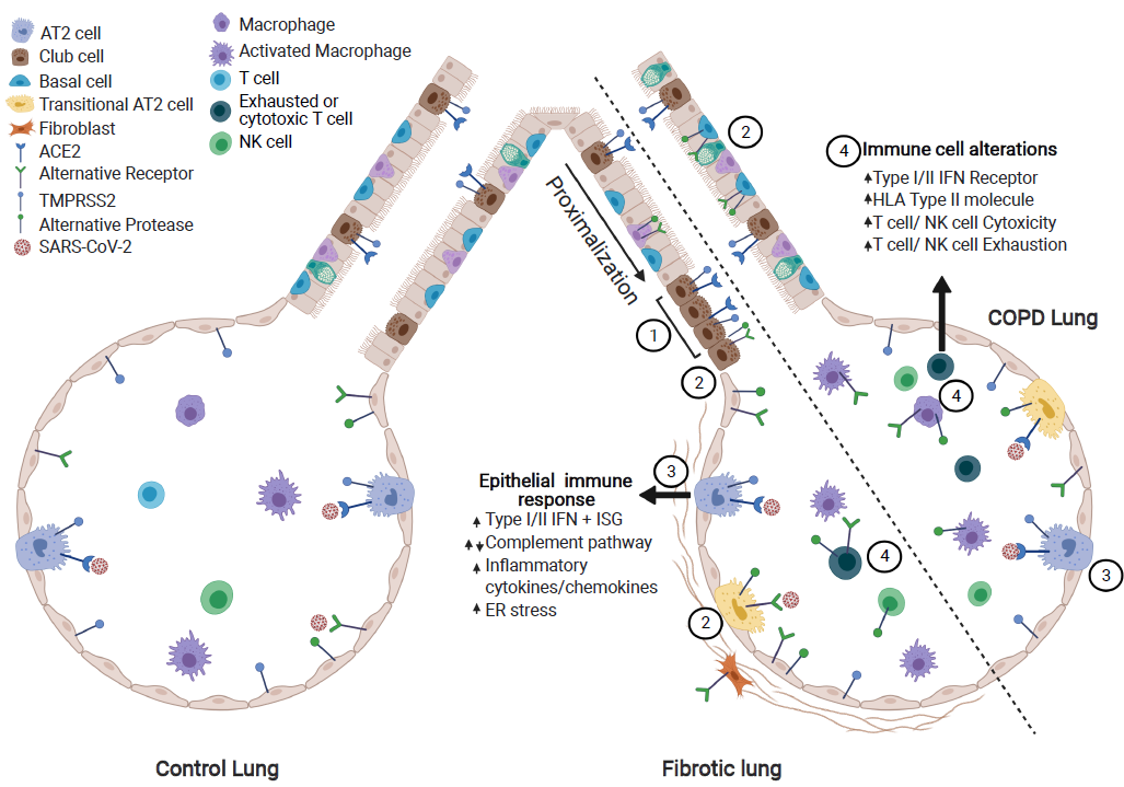
Model of alterations in the diseased lung related to SARS-CoV-2 pathogenesis. (1) In the IPF lung, there is a proximalization of the distal airway. ACE2+ epithelial cells cluster in the small airways though total ACE2+ cell numbers are similar to control. (2) The viral entry score (accounting for all described putative receptors and proteases) is increased in diseased lungs. (3) Diseased epithelial cells have alterations in key SARS-CoV-2 response genes/pathways. (4) In the diseased lung, there is increased expression of cytotoxicity and exhaustion genes in immune cell populations and alterations in viral response pathways (interferon, antigen presentation). Figure created in Biorender.com.
Publications:
Bui L, Winters N, Chung M, et al. 2020. Single-cell RNA-sequencing reveals dysregulation of molecular programs associated with SARS-CoV-2 severity and outcomes in patients with chronic lung disease. bioRxiv 10.20.347187. doi: 10.1101/2020.10.20.347187. Preprint.
Schupp J, Khanal S, Gomez J, et al. 2020. Single-cell transcriptional archetypes of airway inflammation in cystic fibrosis. American Journal of Respiratory and Critical Care Medicine 202(10):1419-1429. doi: 10.1164/rccm.202004-0991OC.
Sungnak W, Huang N, Bécavin C, et al. 2020. SARS-CoV-2 entry factors are highly expressed in nasal epithelial cells together with innate immune genes.Nature Medicine 26(5):681-687. doi: 0.1038/s41591-020-0868-6.
Links:
Abstracts for Dr. Kropski and Dr. Banovich
Public and Technical Abstracts: Integrated Molecular Pathogenesis of Pulmonary Fibrosis
Abstracts for Dr. Schupp
Last updated Thursday, November 13, 2025