Peer Reviewed Cancer


Gold-Containing Nucleosides as Noninvasive Ablation Agents
Posted March 20, 2012
Anthony Berdis, Ph.D., Case Western Reserve University, Cleveland, OH
About 12 million people are affected with some type of cancer. Radiation therapy (RT) is an effective cancer treatment along with surgery and chemotherapy; at least 50 percent of cancer patients receive RT at some point during their cancer treatment. RT uses ionizing radiation (IR) such as x-rays to damage deoxyribonucleic acid (DNA) or genetic material within the tumor cells leading to cell death. However, most IR methods are not specific to cancer cells; i.e., the procedure may also damage normal cells or transform them into cancer cells as the damaged DNA gets inappropriately processed. Therefore, as with other cancer therapies, there are side effects associated with RT.
In an effort to optimize and potentiate RT by combining it with radiosenstizing compounds , Dr. Anthony Berdis received a Fiscal Year 2009 Peer Reviewed Cancer Research Program Concept Award to develop a novel class of chemosensitizer for IR by introducing gold (I) into sulfur-containing nucleosides, an anti-leukemia agent. Attaching gold (I) to a nucleoside analog improves selectivity and efficacy of IR therapy in several significant ways:
- As a biocompatible metal, gold (I) can have potent anti-cancer effects.
- Attachment of nucleoside with gold (I) improves specificity towards cancer cells in comparison to non-cancer cells by bringing the compound directly to cancer cells.
- Gold(I) containing nucleosides also produces beneficial pharmacological effects by targeting kinases (enzymes catalyzing phosphorylation of substrates).
- Reactivity of gold-carbon and gold-sulfur bonds towards radiation amplifies cytotoxity of radiation and improves efficacy of IR therapy.
Dr. Berdis and colleagues synthesized and tested several gold (I) containing compounds on different cancer cells lines such as HeLa (ovarian) and HCT116 (colon) cancer cells. They determined two compounds (labeled as compound 3 and compound 4) hold promising capability to increase the cancer-cell killing activity of IR. The cytotoxity of IR is due to the inability of cancer cells to repair double-strand DNA breaks (DSBs), the most lethal form of DNA damage caused by IR. HeLa cells treated with RT alone showed a survival curve reflecting cells capable of repairing damaged DNA created by the IR exposure. However, in cells treated with either compound 3 or compound 4 for 24 hours followed by RT, the repairing phase is absent, indicating an increase in the cytotoxicity of IR treatment.
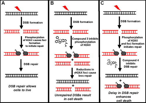
Dr. Berdis also elucidated the cellular mechanism for potentiating the anti-cancer effects of the gold (I) containing compounds. H2AX is a protein which gets phosphorylated and activated in response to DSBs formed by IR exposure. As the figure demonstrates, in the absence of compound 3, H2AX gets phosphorylated to repair DSB, while in the presence of compound 3, H2AX phosphorylation is inhibited. The DSBs are thus not repaired, resulting in cell death. On the other hand, compound 4 treatment inhibits DSBs repair process in the step after activation of H2AX.
Therefore, the accumulative results of this proof-of-concept study suggest combination of gold (I) containing compounds with IR, as a potent therapeutic strategy for tumor ablation, may have a major impact on cancer research. Dr. Berdis continues to pursue these studies with more advanced experiments to move the combination therapy of gold (I)-containing compounds and IR chemostrategy forward.
Schematic illustration showing proposed model of gold (I) containing nucleosides to potentiate the therapeutic effects of IR by inhibiting DNA repair