


Posted September 4, 2015
Dr. David Karow, MD, Ph.D., University of California, San Diego
PC120532
 Technology is improving our ability to detect prostate cancer, but is still unable to reliably determine which patients have aggressive disease that requires immediate treatment, and which have low grade indolent tumors that can be safely monitored and perhaps forego treatment all together. Current diagnostic methods rely on invasive collection of biopsy samples for pathological analysis, so researchers are looking for non-invasive methods that can reliably distinguish between aggressive and indolent disease.
With support from a FY12 New Investigator - Idea Development Award, Dr. David Karow of the University of California, San Diego (UCSD) implemented a new technique, called Restriction Spectrum Imaging (RSI), which utilizes Magnetic Resonance Imaging (MRI) to non-invasively detect and distinguish aggressive from indolent prostate cancer. This technique was originally pioneered in the brain by UCSD Radiology faculty Dr. Anders Dale, PhD and Dr. Nate White, PhD for the detection and characterization of brain tumors. RSI measures the restricted diffusion of water within a cell, which differs between normal prostate tissue and cancerous tissue due to alterations in cell morphology. These seemingly subtle differences are measureable by MRI scanners and have been found to be highly correlated with the pathologically determined Gleason grades measured in biopsy samples as assessed by Dr. Ahmed Shabaik, MD, Professor in the UCSD Department of Pathology. In addition, in work recently submitted for publication, Dr. Karow and his team have found that a rapid five-ten minute exam which includes RSI is equally capable of detecting high grade disease as a full 60 minute contrast-enhanced, multi-parametric MRI. Overall, Dr. Karow's findings support the potential for RSI as a non-invasive, rapid in vivo biomarker for prostate cancer detection and characterization.
Working with UCSD Urology faculty members, Dr. Chris Kane, MD and Dr. J. Kellogg Parsons, MD, MHS, Dr. Karow and his team have transitioned this new method into clinical practice. RSI is utilized on all patients imaged at the UCSD medical center, and they are actively engaged with other select institutions to make this imaging technique more widely available. In addition, Dr. Karow and his team have expanded the applications of RSI to include improved direction of targeted procedures including biopsy collection.
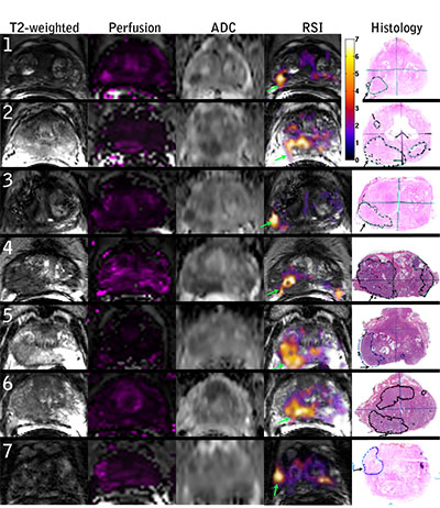
MR imaging and whole mount pathology for 7 prostate cancer patients. Column 1: T2 - weighted images, column 2: Perfusion Ktrans maps, column 3: standard apparent diffusion coefficient (ADC) maps (b = 0, 100, 400, and 800 s/mm2), column 4: RSI z-score maps, column 5: whole mount pathology with tumor and area of EPE identified. Color bar represents z-scores from 0 to 7 for the RSI maps. (Rakow-Penner et al. Novel Technique for Characterizing Prostate Cancer Utilizing MRI Restriction Spectrum Imaging: Proof of Principle and Initial Clinical Experience with Extraprostatic Extension. Prostate Cancer Prostatic Dis. 2015 March; 18(1):81-5)
Research Links:
Links: