


Posted April 15, 2014
Dr. Wubao Wang, City University of New York
 Physicians routinely use two examinations to screen for prostate cancer: the prostate specific antigen (PSA) blood test and a digital rectal examination (DRE). An abnormal DRE and an elevated PSA level are both possible indicators of the disease; however, neither test by itself or in combination can provide a definitive diagnosis of prostate cancer. If either test is abnormal, physicians will usually perform an invasive biopsy procedure, which is painful and damaging to prostate and other tissues. Therefore researchers are working to develop new diagnosis methods that will be both less invasive and more accurate.
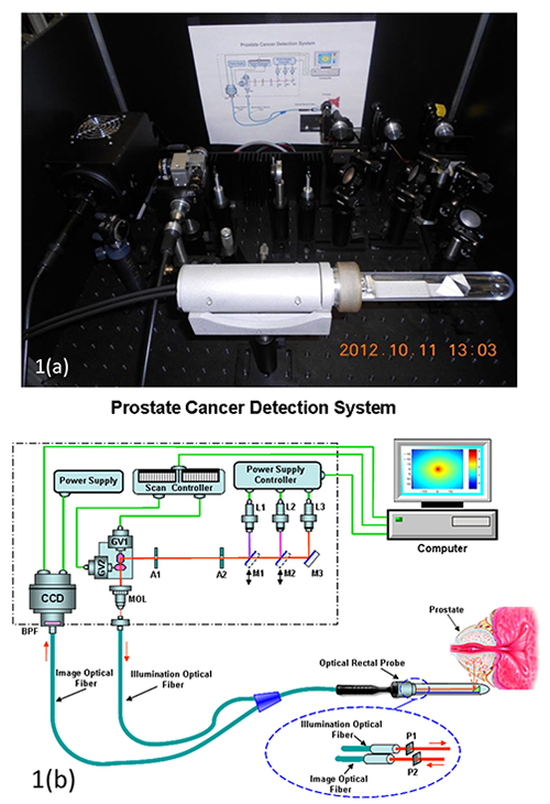
Dr. Wubao Wang's group at the City University of New York (CUNY) has been working to develop a less invasive imaging technique with support from a 2007 PCRP Idea Development Award. Says Dr. Wang, "The key feature of our rectal scanning imager is the use of near infrared (NIR) radiation to image the prostate through the rectum. This imaging approach will greatly improve and supplement current (detection) methods of PSA and DRE because the scanning imaging unit and inverse image reconstruction technique can be used to map the internal structure of the prostate, distinguishing cancerous from normal areas."
This group has utilized 2D image acquisition software combined with an inverse image reconstruction algorithm to record sets of 2D images of the prostate in animal models and then subsequently determine the existence and 3D locations of cancerous sites. The group is now preparing to extend this testing to the clinical setting, evaluating the efficacy of rectal scanning imaging for prostate cancer diagnosis before ultrasound and biopsy measurements. In addition, he plans to incorporate the use of "smart" fluorescent dyes to target and mark prostate cancer receptors and cells, providing identification of aggressive metastatic cancers in early stages. These advanced methods for earlier and improved detection of prostate cancer will help physicians provide better prostate cancer management and protect patients with low risk disease from unnecessary treatment.
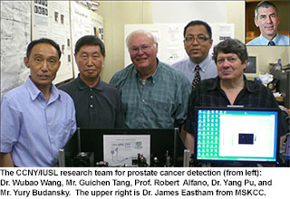
The key investigators in the CUNY research group include Prof. Robert Alfano (Director of the Institute for Ultrafast Spectroscopy and Lasers), Dr. Wubao Wang, and Dr. Yang Pu. The research achievement was made in collaboration with Dr. James Eastham at Memorial Sloan-Kettering Cancer Center, Prof. Samuel Achilefu at Washington University School of Medicine, and Prof. Min Xu at Fairfield University.
Publications:
Yang Pu, Wubao Wang, Min Xu, James A. Eastham, Guichen Tang, and Robert R. Alfano, "Characterization and three-dimensional localization of cancerous prostate tissue using backscattering scanning polarization imaging and independent component analysis", Journal of Biomedical Optics, 17 (8), 081419-1-8 (2012).
Yang Pu, Wubao Wang, Yuanlong Yang, and R. R. Alfano, "Stokes shift spectroscopy highlights differences of cancerous and normal human tissues", Optics Letters, 37(16), 3360-3362 (2012).
Y. Pu, W. B. Wang, G. C. Tang, and R. R. Alfano, "Changes of collagen and NADH contents in human cancerous and normal prostate tissues studied using fluorescence spectroscopy with selective excitation wavelength", Journal of Biomedical Optics, 15, 047008-1-5 (2010).
Links: