


Posted November 6, 2014
M. Albert Thomas, Ph.D., University of California at Los Angeles
 Current methods of prostate cancer (PCa) detection, including digital rectal examination (DRE), serum prostate-specific antigen (PSA) level measurement, and transrectal ultrasound (TRUS) based biopsy, are invasive and in general lack reliable specificity and accuracy. Researchers have therefore been focusing on how to achieve greater sensitivity and specificity to accurately detect prostate cancer through non-invasive imaging techniques. One non-invasive imaging method available on most magnetic resonance imaging (MRI) scanners, proton (1H) magnetic resonance spectroscopy (MRS), can read the chemical content of tissues, such as metabolites. Since cancer cells have higher growth rates than normal cells, measurement of changes in metabolite concentrations might help detect the presence of cancer cells, and even distinguish between rapidly growing, aggressive cancer cells and indolent cancer cells.
Current one dimensional (1D) MRS detection of metabolites is limited because of signal overlap, making the measurement of individual metabolites associated with cancer growth difficult to tease apart. With support from a 2010 PCRP Idea Development Award, Dr. M. Albert Thomas and his research team at the University of California at Los Angeles set out to develop methods for the acquisition of multi-dimensional 1H MRS scans to better resolve measurement of metabolites for prostate cancer diagnosis. His group has demonstrated a powerful noninvasive technique called Echo-Planar J-resolved Spectroscopic Imaging (EP-JRESI) that can distinguish cancerous tissue from healthy tissue (Figure 1). Dr. Thomas showed that multi-dimensional MRS detection, which allows resonance signals to be spread apart, allows for the quantitation of several metabolites whose signal were overlapped in the 1D spectrum. Analysis of several molecules simultaneously improves the detection and diagnosis potential of 1H MRS, allowing researchers and physicians to get a better grasp on the metabolism of prostate regions and ultimately detect aggressive disease.
Dr. Thomas' novel 4D EP-JRESI technique has been implemented on 3T and 1.5T MRI scanners, and a pilot study of PCa patients has been completed, with follow up studies planned to establish clinical feasibility. Additional plans are in development to establish a novel protocol for multi-parametric MR examination combining the 4D EP-JRESI (metabolite imaging) with anatomical MRI (T2-weighted) and functional MRI. It is Dr. Thomas' hope that the recent developments will lead to clinical realization of these novel multidimensional magnetic resonance spectroscopy imaging (MRSI) sequences in the near future for improved detection and diagnosis of PCa.
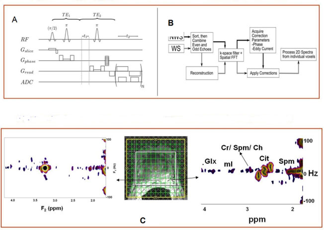
A) A schematic diagram of the 4D EP-JRESI sequence implemented on the MRI scanner.
B) Processing algorithm logic flow
C) MRI & MRS spectra from a 61- year old PCa patient (left) normal (right) cancerous tissues
Research Links:
1) Thomas MA, Nagarajan R, Huda A, Margolis D, Sarma MK, Sheng K, Reiter RE, Raman SS. "Multidimensional MR Spectroscopic imaging of Prostate Cancer In vivo." NMR in Biomed 2014; 27:53-66; Epub 2013 July 31. [PMID: 23904127].
2) Furuyama J, Wilson NE, Burns BL, Nagarajan R, Margolis DJ, Thomas MA. "Application of Compressed Sensing to Multidimensional Spectroscopic Imaging in Human Prostate." Magn Reson Med 2012; 67:1499-1505 [PMID: 22505247]
3) Nagarajan R, Margolis D, Raman S, Sarma MK, Sheng K, King CR, Verma G, Sayre J, Reiter RE, Thomas MA. "MR Spectroscopic Imaging and Diffusion-Weighted Imaging of Prostate Cancer with Gleason Scores." J Magn Reson Imaging 2012;36:697-703 [PMID: 22581787]
4) Additional Publications
Links: