


Posted August 6, 2013
Rohit Bhargava, Ph.D., University of Illinois
 The presence of prostate cancer is usually first suspected through a PSA test result or a physical examination. Men who are suspected to have prostate cancer undergo a prostate biopsy, and the biopsy specimens are analyzed by a pathologist who examines the tissue under a microscope and identifies certain characteristics of the cells and tissue architecture that indicate the presence and potential severity of cancer. However, this long-established method of recognizing disease and predicting its progression is an imperfect science and subject to some interpretation, and misdiagnosis can put some patients at risk for allowing their cancer to develop to aggressive disease or cause other patients to undergo unnecessary treatment for tumors that may never harm them.
At the University of Illinois, Professor Rohit Bhargava has developed a novel form of microscopy that not only measures tissue structure, but also the chemical content of every cell. With support from his FY06 PCRP New Investigator Award, he was able to apply this method, termed infrared spectroscopic imaging, to analyze prostate tissue biopsies. Working with a team of engineers, surgeons, and pathologists, Dr. Bhargava developed sophisticated mathematical algorithms that allow him to look for cell types that determine the presence and severity of cancer with an accuracy that is unmatched by current methods. By looking at the chemical changes in each cell type present in the prostate tissue, this technique identified changes in specific cell types that correlated with a higher probability of recurrence, thereby identifying which patients would be more likely to develop advanced disease after treatment. These findings have now been validated on a large scale, and Dr. Bhargava plans to bring this new technique to multi-site clinical trials. Successful translation to the clinic could provide major benefits for prostate cancer patients in the near-term future.
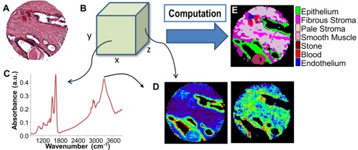
(A) Conventional imaging in pathology requires dyes and a human to recognize cells. In chemical imaging data cubes (B), both a spectrum at any pixel (C) and the spatial distribution of any spectral feature can be seen, e.g. in (D) nucleic acids (left, at ~1080 cm-1), and collagen specific (right, at ~1245 cm-1). Computational tools can then convert chemical imaging data to knowledge used in pathology (E).