 An official website of the United States government
 ) or https:// means you've safely connected to the .mil website. Share sensitive information only on official, secure websites.
Orthotics and Prosthetics Outcomes


Posted February 24, 2023
Jeanne Bailey, Ph.D. and Richard O'Donnell, M.D., University of California, San Francisco

Dr. Jeanne Bailey
(Photo Provided)
Osseointegration is a newer technology available in the United States to help individuals with lower limb and upper limb amputation. Traditionally, a socket-interface is created to physically attach a prosthesis to the residual limb, for example, via a prosthetic liner suspension system. Osseointegration involves surgically implanting a bone-anchor into the base of the remaining, or residual, limb. This implant extends from the limb and directly connects to a prosthesis. Osseointegration provides advantages over the traditional prosthetic socket as it eliminates the need for the socket, which often causes skin breakdown, pain, and discomfort. Approximately 7.4% of U.S. military personnel underwent major limb amputation due to causalities sustained during conflicts from 2001-2006 (Stansbury et al. 2008). As the number of Veterans and patients seeking and undergoing osseointegration procedures worldwide increases, more data is needed to understand the advantages and effects of this option compared to traditional prosthetic socket technology.
Due to altered gait mechanics and joint loading patterns, transfemoral amputees are at risk for developing hip joint degradation conditions, which can increase the likelihood of needing a future hip replacement. The osseointegration bone implant system could likely impact the ability to complete a total hip replacement due to the limited room along the shaft of the femur to contain both the osseointegrated implant and hip replacement hardware. At the University of California, San Francisco (UCSF), Drs. Jeanne Bailey and Richard O'Donnell are investigating how the hip muscles support the residual limb in the traditional prosthetic socket technology compared with the osseointegration technology to better understand outcomes associated with available treatment options.
With funding from a Fiscal Year 2019 (FY19) Orthotics and Prosthetics Outcomes Research Program (OPORP) Clinical Research Award, Drs. Bailey and O'Donnell are assessing biomechanical function and hip-stabilizing muscle quality associated with transfemoral osseointegration. This award builds upon their initial Department of Defense-funded pilot study. Dr. Richard O'Donnell's Osseointegration Clinic at UCSF was the first civilian center in the United States to specialize in the use of percutaneous osseo-anchored titanium implants for the rehabilitation of patients with amputation.
The FY19 OPORP-funded research project is a cohort study that compares outcomes of unilateral transfemoral osseointegrated amputees with healthy controls. The osseointegrated amputees will be at least 2 years from final surgery. Dr. Bailey's research team will capture biomechanical data to quantify movement quality during walking and sit-to-stand actions. Additionally, magnetic resonance imaging (MRI) scans will be collected from these same participants to glean more information regarding the quality and characteristics of their hip muscles.
Preliminary data demonstrates that osseointegrated patients appear to be losing muscle quality (more muscle fat infiltration) after surgery (Figure 1). But it is unclear how this asymmetry in muscle quality affects biomechanical function. The next step of the team's analysis will explore the association between hip muscle quality and biomechanical function affecting gait for lower extremity amputees. This, in turn, can help improve targeted rehabilitation for improving hip muscle quality and long-term outcomes. The data also supports the addition of an MRI assessment of muscle quality and lower extremity joint health to the comprehensive care plan for amputees.
Taken together, results from this study will improve osseointegration surgical outcomes and provides recommendations for interpretation of long-term biomechanical outcomes in these patients. Overall, this study stands to identify long-term risk factors associated with the implant and strategies for mitigating risk for gait dysfunction and hip osteoarthritis following transfemoral amputation due to poor hip-stabilizing muscle quality.
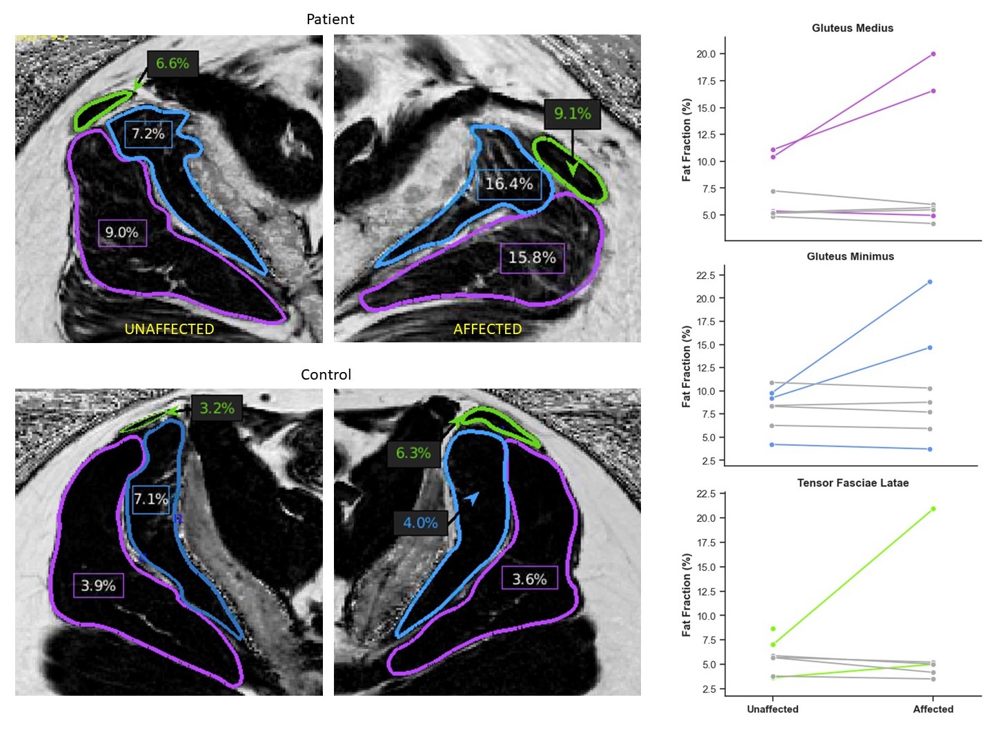 Figure 1: Fat percent for hip muscles; Gluteus Medius (purple), Gluteus Minimus (blue), and Tensor Fasciae Latae (green). The image on top provides an IDEAL MRI of a patient with an osseointegrated prosthesis versus a side matched control below comparing both amputated (affected) and unaffected sides. (Figure Provided)
References:
Stansbury LG, Lalliss SJ, Branstetter JG, et al. 2008. Amputations in U.S. military personnel in the current conflicts in Afghanistan and Iraq. Journal of Orthopaedic Trauma
Last updated Monday, September 22, 2025