
Ovarian Cancer
Optical Imaging Falloposcope for Minimally Invasive Ovarian Cancer Detection


Posted September 27, 2016

Dr. Jennifer Barton
(University of Arizona)
(University of Arizona)
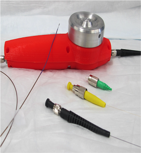
Prototype falloposcope, showing handle (red), optical connections (black, yellow, green), and the 0.8mm diameter imaging tip. (Courtesy of Dr. Barton).
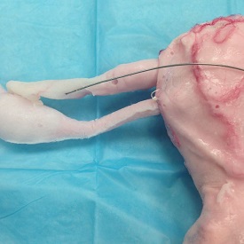
Imaging tip of falloposcope prototype next to silicone model of the female reproductive tract (Courtesy of Dr. Barton)
Dr. Jennifer Barton, recipient of an FY12 OCRP Pilot Award, has developed and patented a new ovarian cancer screening device which she hopes can be used to detect this cancer early and save many lives. Ovarian cancer is one of the deadliest forms of cancer today, primarily because there are no effective or widely used tests to detect it at its earliest stages when it is most treatable. There is no doubt that widespread screening practices for cancers of the breast, cervix, and colon have extended and even saved many lives, and the success of Dr. Barton's new Falloposcope technology represents a huge opportunity to do the same for ovarian cancer. Although the overall 5-year survival rate for ovarian cancer patients is only 46%, the rate for patients whose cancer is discovered when it is still confined to the ovary is over 90%. Unfortunately, only 15% of cases of are diagnosed at this local, treatable stage¹.
Dr. Barton's concept is simple: the falloposcope is designed to be used as part of a minimally invasive outpatient screening procedure, similar to colonoscopy. The falloposcope consists of two highly sensitive imaging systems mounted on an endoscope that is less than 1 millimeter (mm) in diameter. By combining two imaging systems, called optical coherence tomography (OCT) and multispectral fluorescence imaging (MFI), doctors can assess both how the tissue looks and how it functions, allowing them to distinguish between normal, cancerous, and benign tissue. Having overcome the engineering challenges involved in designing the optics and mechanics of the systems within the confines of a 0.8 mm diameter tip, the falloposcope components have been fabricated and assembled.
To date, the falloposcope has been tested in a silicone surgical simulation model of the human uterus, fallopian tubes, and ovary, which was developed by Dr. Barton’s team at the University of Arizona. In that model, which has optical and mechanical properties similar to human tissue, the mechanical performance and image quality delivered by the falloposcope have been excellent. The device has also been tested in a pilot study of 47 ovarian and 31 fallopian tube tissue samples from women who underwent oophorectomy or debulking surgery. In that setting, MFI/OCT imaging was able to identify disease in both fallopian tubes and ovaries, and benign disease was distinguishable from cancer.
Dr. Barton envisions this technology first being used in women at high risk for ovarian cancer, enabling them to delay or avoid prophylactic oophorectomy. A full patent has been filed on this technology, and it is currently being marketed for license. If proven effective, the falloposcope might be used to screen and save women's lives.
Links:
¹ American Cancer Society, Cancer Facts & Figures 2016
Last updated Friday, December 13, 2024