


Posted March 8, 2017
Seth Smith, Ph.D.
Vanderbilt University Medical Center
Dr. Seth Smith (far right) and team
Vanderbilt University Medical Center
Vanderbilt University Medical Center
Dr. Seth Smith received a fiscal year 2012 (FY12) Idea Award from the Multiple Sclerosis Research Program (MSRP) to develop new magnetic resonance imaging (MRI) tools that can assess gray matter changes in multiple sclerosis (MS) patients and correlate those changes to cognitive function. To tackle this challenge, Dr. Smith used 7T, MRI which has more than twice the strength of many conventional MRI units. This allowed him to examine the brain structures in finer detail than what can be achieved with typical clinical 3T MRI units. The fact that he chose to study gray matter is also a unique feature of this project; most studies of MS focus on the white matter because often the gray matter cannot be as easily described with the currently available clinical MRI tools.
For this study, Dr. Smith and his group focused on three types of gray matter damage: loss of myelin, change in tissue biochemistry, and change in tissue function. The umbrella research question was: can an advanced set of high-field (7T) MRI tools offer insight into a patient's cognitive decline in MS? Dr. Smith's goal was to show that there is a relationship between 7T MRI findings and a patient's ability to focus or remember. Thus, he felt that looking at what is going on macroscopically (myelin), microscopically (biochemical), and functionally, would allow him and his team to unravel the changes that occur in a MS patient's gray matter that result in cognitive impairment.
To this end, Dr. Smith and his team have developed a toolbox of quantitative MRI methods that show definitive differences between MS patients and healthy volunteers. Specifically, three of the methods developed using 7T MRI assessed markers that correlated well to patient cognitive function. In addition to having success using the more powerful, yet less clinically relevant, technology, Dr. Smith was also able to develop and deploy two of the three methods to the more clinically accessible 3T MRI. The adapted methods have now been used not only in this study, but by other clinicians at other institutions and even in other countries.
Dr. Smith is particularly proud of the strength of his team, where a person from nearly every aspect of research came together to ensure the success of this project. During the course of the award, the team included an undergraduate student, two research assistants, a graduate neuroscience Ph.D. student, a postdoctoral fellow, a MS neuroimmunologist clinician, a neuroradiology resident, and Dr. Smith- all making important contributions to this work. Dr. Smith was "especially encouraged by this [team] as it shows the impact that can be had when communities of researchers gather and learn from each other."
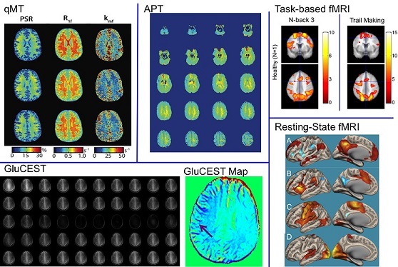
Example data for quantitative Magnetization Transfer (qMT) (PSR, R1f, kmf), Amide Proton Transfer (APT asymmetry), Task-based fMRI (NBack, Trail Making), Glutamate Chemical Exchange Saturation Transfer (GluCEST), and Resting-state fMRI for one healthy volunteer showing the range of data acquired.
Publication:
Dula AN, Pawate S, Dortch RD, et al. 2015. Magnetic resonance imaging of the cervical spinal cord in multiple sclerosis at 7T. Mult Scler. 22(3):320-328. DOI: 10.1177/ 1352458515591070.
Link:
Last updated Monday, March 10, 2025