 An official website of the United States government
 ) or https:// means you've safely connected to the .mil website. Share sensitive information only on official, secure websites.
Epilepsy
Large Animal Models of TBI May Reveal the Mechanisms Behind Post-Traumatic Epilepsy


Posted December 22, 2020
John A. Wolf, Ph.D., University of Pennsylvania
 Dr. John A. Wolf
Many Service members and civilians who suffer a TBI later develop spontaneous recurrent seizures, i.e., PTE. During the time between the TBI and the development of PTE, the brain undergoes changes that make it prone to seizures, a process known as epileptogenesis. The mechanisms of epileptogenesis are not fully understood, and predictors (biomarkers) of who will go on to develop PTE following a TBI are not well established. Both of these questions, how to intervene and on which TBI patients to deploy an intervention, are the goal of a new study.
Dr. John Wolf, of the University of Pennsylvania and the Philadelphia CMC VA Medical Center, is using a large animal (pig) model to break new ground in PTE research. This model of PTE approximates the human brain better than a rodent, and allows Dr. Wolf to measure the degree to which different injury types contribute to the development of epilepsy. He has also carefully characterized the extent of the injuries to these animals in an effort to understand which brain structures are most susceptible to damage from a head injury, as well as which regions might become hyperexcitable or dysfunctional leading to acquired epilepsy. In addition, measurements from the blood are being taken in order to predict which animals will go on to develop epilepsy in hopes of using this information to make better predictions for patients.
Another major challenge that Dr. Wolf is addressing is how to measure alterations in brain activity after injury over long periods of time, which has never been done previously in a large animal model. Dr. Wolf and his team designed an integrated system that can measure the spread of abnormal electrical activity in the brain after injury, including in deep brain structures with high resolution. The preliminary data generated by this work has already led to a greater understanding of how early post-injury dysfunctional activity occurs. Dr. Wolf is preparing to generate maps of how adjacent regions of the brain alter their communication after injury. Understanding the start and spread of these network disruptions will reveal novel mechanisms related to epileptogenesis and may lead to advances in seizure detection, as these experiments may reveal the earliest events and locations involved in PTE. A better understanding of the mechanisms of epileptogenesis should allow for more targeted treatments, with the goal of blocking the development of epilepsy following TBI.
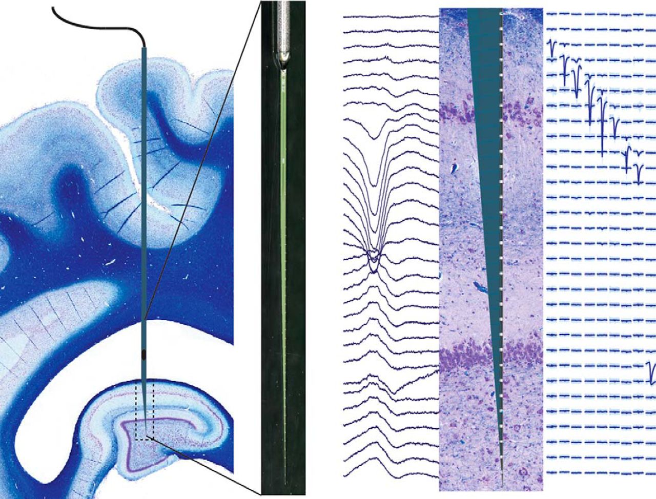 Histological characterization of the dorsal hippocampus of Yucatan miniature pigs. Stereotaxis combined with single-unit electrophysiological mapping was used to precisely place multichannel laminar silicon probes into the dorsal hippocampus without the need for image guidance. In vivo electrophysiological recordings of simultaneous laminar field potentials and single-unit activity in multiple layers of the dorsal hippocampus were used to physiologically identify and quantify these layers under anesthesia.
Reference:
Ulyanova A, Koch P, Cottone C, et al. 2018. Electrophysiological signature reveals laminar structure of the porcine hippocampus. eNeuro 5(5):ENEURO.0102-18. DOI:10.1523.
Link:
Last updated Thursday, November 13, 2025