
Military Operational Medicine (JPC-5)
Identifying Biomarkers that Distinguish Post-Traumatic Stress Disorder and Mild Traumatic Brain Injury Using Advanced Magnetic Resonance Spectroscopy


Posted June 26, 2020
Alexander Lin, Ph.D., Brigham and Women's Hospital, Inc., and John M. Irvine, Ph.D., The Charles Stark Draper Laboratory, Inc.

Dr. Alexander Lin

Dr. John Irvine
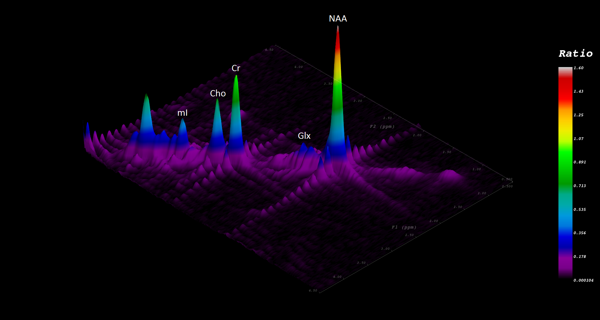
Mild traumatic brain injury (mTBI) and post-traumatic stress disorder (PTSD) are two highly prevalent injuries sustained by U.S. military forces deployed in service of Operation Enduring Freedom and Operation Iraqi Freedom. Distinguishing the two can be difficult due to overlapping and transient symptoms. Additionally, current diagnostic techniques rely largely on subjective measures and self-report questionnaires. Drs. Alexander Lin and John Irvine proposed to address this gap by developing objective diagnostic criteria using a noninvasive neuroimaging technology, magnetic resonance spectroscopy (MRS). MRS can detect various chemical changes in the brain and has the potential to enable accurate diagnosis of neurological disorders such as Alzheimer’s disease. Building upon conventional 1D MRS techniques, 2D correlation spectroscopy (COSY) capitalizes on the molecular signals, or resonances, that are coupled resulting in the ability to distinguish different molecular signals in higher-resolution data. COSY spectroscopy in homogenous samples is common in biological and chemical studies. However, COSY spectroscopy of heterogeneous samples, such as those of the living tissue environment, is much more complex. The utility of COSY in living organisms is less prevalent due to increased scan times and lack of accessible, standardized data processing methods. Addressing scan times and data processing challenges may allow MRS techniques such as COSY to become more widely used in diagnostic radiology. Drs. Lin and Irvine received a Fiscal Year 2009 Investigator-Initiated Research Award through the Psychological Health and Traumatic Brain Injury Research Program to use MRS methods, such as COSY, and classification algorithms to be able to detect and distinguish mTBI and PTSD based on objective measures of changes in brain biochemistry using 1D and 2D MRS methods.
The researchers developed, tested, and implemented a processing pipeline for harmonizing 1D and 2D MRS data from multiple sources and identified robust biomarkers that differentiate MRS signals in civilians, Soldiers with mTBI, and Soldiers with PTSD. In addition to correlating their findings with functional outcomes, standardized protocols from this study were then corroborated in a study of retired National Football League players and have been disseminated through the Enhanced NeuroImaging Genetics through MetaAnalysis (ENIGMA) network.
This study recruited five groups of subjects for MRS data collection: (1) Soldiers with PTSD but not mTBI; (2) Soldiers with mTBI but not PTSD; (3) Soldiers with both mTBI and PTSD; (4) pre-deployment Soldiers without history of mTBI or PTSD (military control); and (5) civilians without history of mTBI or PTSD (civilian control). Data from 1D MRS experiments informed improvements of biomarker identification algorithms, which were then incorporated into 2D MRS analyses. 2D COSY data required analysis by machine learning algorithms, which were trained using features identified by an independent component analysis. The researchers then developed a 2D COSY processing pipeline that addressed signal drift, low signal-to-noise ratios, dominating water signals, lipid contamination, and inter-subject alignment of spectra to landmark peaks. The identified classifiers and biomarkers for PTSD and mTBI were then tested for performance and robustness with additional data.
The proposed classifiers and biomarkers were based on the ratios of several neurochemicals (e.g., creatine, glutamate, glutathione, myo-inositiol) in different regions of the brain (e.g., posterior cingulate gyrus, parietal white matter) in each group of subjects. In their analyses, the researchers found statistically significant markers that differentiated military controls and civilian controls, as well as markers for those with mTBI only or PTSD only compared to those with mTBI and PTSD diagnoses. Further, they correlated some of those markers with clinical cognitive performance (including processing speed and verbal memory) and disseminated standardized protocols through the ENIGMA network. Ultimately, these findings require full validation in additional studies to confirm their performance and candidacy for diagnostic biomarkers. Diagnostic criteria to differentiate mTBI and PTSD will be profoundly beneficial for return-to-duty decision making, delivery of timely and appropriate treatment and rehabilitation services to affected Service members, as well as long-term health and well-being of Service members and Veterans.
Publications:
Miller MW, Lin AP, Wolf EJ, and Miller DR. 2018. Oxidative stress, inflammation, and neuroprogression in chronic PTSD. Harvard Review of Psychiatry 26(2):57-69.
Brix KA, Brody DL, Grimes JB, and Yitzhak A. 2017. Military blast exposure and chronic neurodegeneration: Summary of working groups and expert panel findings and recommendations. Journal of Neurotrauma 34(S1):S18-S25.
Rowland B, Liao HJ, Adan F, et al. 2017. Correcting for frequency drift in clinical brain MR spectroscopy. Journal of Neuroimaging 27(1):23-28.
Wilde EA, Bouix S, Tate DF, et al. 2015. Advanced neuroimaging applied to Veterans and service personnel with traumatic brain injury: State of the art and potential benefits. Brain Imaging and Behavior 9(3):367-402.
Cocuzzo D, Lin A, Stanwell P, et al. 2014. In vivo brain magnetic resonance spectroscopy: A measurement of biomarker sensitivity to post-processing algorithms. IEEE Journal of Translation Engineering in Health and Medicine 2:1-17.
Ng TS, Lin AP, Koerte IK, et al. 2014. Neuroimaging in repetitive brain trauma. Alzheimer's Research and Therapy. 6(1):10.
Ramadan S, Lin AP, and Stanwell P. 2013. Glutamate and glutamine: A review of in vivo MRS in the human brain. NMR in Biomedicine 26(12):1630-1646.
Link:
Last updated Friday, March 7, 2025