
Defense Medical Research and Development
Army Medicine Opens the Door for Vision Prosthetic Prototypes


Army medical research funding has supported the development of innovative technologies to assist injured Service members and improve their quality of life. For example, advances in prosthetics for those with loss of limbs have enabled them to regain independence in work, sports, and in their daily lives. In contrast, restoring vision has been more challenging.
In May 2014, experts in various science, technology, and regulatory disciplines from the Government, industry and academia were brought together by the US Army Medical Research and Materiel Command (USAMRMC) to evaluate the maturity of visual prosthesis technology. The group consensus on the readiness for prototyping within this area was, “Yes!” As a first step, the Clinical and Rehabilitative Medicine Research Program, on behalf of the USAMRMC, released the Vision Prosthesis Pilot Study Award funding opportunity in 2015. This initiative sought to fund research exploring novel technologies contributing to the development of a working visual prosthesis prototype for individuals who have sustained severe macular degenerate or traumatic eye injury. The USAMRMC is now announcing the three successful awardees.
“When the eye and optic nerve are severely damaged, the hope of a cortical visual prosthetic is that by directly stimulating the brain’s visual cortex, it can restore sight,” said Dr. Kenneth Bertram, Principal Assistant for Acquisition, USAMRMC. We are very pleased that the initial funding brings us three different approaches to the development of visual prostheses.”
Skipping the damaged optic nerve:
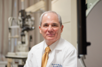
Joseph F. Rizzo, M.D., Massachusetts Eye and Ear, Harvard Medical School
Dr. Joseph Rizzo’s team, from Massachusetts Eye and Ear, has been a leader in this research through the collaboration of Harvard Medical School and the Massachusetts Institute of Technology on the Boston Retinal Implant Project (BRIP). For those with severe injury in both eyes, as sometimes occurs with blast injuries, both the eyes and the optic nerves may be damaged. As a result, making the connection of an image from the prosthetic eye to the brain is a challenge. The BRIP group has developed a wireless and implantable neural prosthesis for use in the retina. However, this device relies on a functional optic nerve. With this award Dr. Rizzo’s team will develop and test a prosthesis to electrically stimulate the lateral geniculate nucleus — a key relay center in the brain’s visual processing pathway — that will allow signals to skip over the damaged optic nerve and reach visual centers in the brain.
Cleaning up that image:
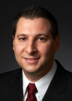
Andrew C. Weitz, Ph.D., University of Southern California, Roski Eye Institute
Dr. Andrew Weitz and his team from University of Southern California, Roski Eye Institute, have focused on the difficulty of generating precise images with a cortical visual prosthesis. In a cortical visual prosthesis, cameras capture patterns of light and relay those patterns to electrodes implanted in the brain’s visual cortex. The electrodes deliver current to nerve cells in the visual cortex, causing them to fire and create an image in the subject’s visual field. A key problem in this approach is that in addition to stimulating the target nerve cells, electrodes can also stimulate passing nerve axons that lead to more distant visual cortex nerve cells, causing them to also fire. The result can be an inaccurate image polluted by multiple spots of light appearing all over the patient’s visual field. To attack this problem, Dr. Weitz’s team developed a novel fluorescence imaging technique to map the patterns of cells that are activated by electrical stimulation. Using this technique in experiments in the retina, Dr. Weitz’s team identified varying electrode stimulus waveform shapes that avoid axons and confine the retinal activation to the area around the electrode. Using funding from this award, the team is now developing waveforms that will enable them to repeat this success in the visual cortex. This will guide the design of microelectronic arrays and stimulus generator hardware for use in a prototype cortical visual prosthesis. The prediction is that this cortical visual prosthesis will produce visual field images that better match the world in front of the subject.
Doing it without electrodes:

Joseph P. Kao, Ph.D.,
University of Maryland,
School of Medicine Center for Biomedical Engineering & Technology
Dr. Joseph Kao, professor of physiology at University of Maryland School of Medicine Center for Biomedical Engineering & Technology (BioMET), is exploring the use of optical photostimulation — using focused light flashes to photo-release neurotransmitter molecules that stimulate neurons in the brain’s visual cortex. Normally, light coming in through the eye stimulates cells that send signals back through the optic nerve, and on through the brain, to release chemicals called neurotransmitters. The neurotransmitter turns on cells in the visual cortex, allowing us to see. Instead of implanting electrodes into the visual cortex, Dr. Kao with colleagues from BioMET and University of Maryland, College Park, plan to synthesize a neurotransmitter molecule whose activity is blocked by a photosensitive “cage.” Exposure to a flash of light breaks the cage to unleash the fully active neurotransmitter. The team will test the ability of a small light-emitting device placed on the surface of the brain to activate caged neurotransmitter deeper in the visual cortex, activating neurons and producing an image. Developing and validating a technology based on photo-releasable neurotransmitters could enable a less invasive approach for vision prosthetics.
“This is only the beginning,” said Dr. Bertram, “…some obstacles and direction will be found through the studies of these prototypes. Our initiative is to restore vision and improve the quality of life for our wounded Warriors and other Americans who have become blind.”
Dr. Rizzo's photo by John Earle, and Drs. Weitz and Kao photos were courtesy of their institutions.
Links:
Defense Medical Research and Development Program
Last updated Thursday, December 5, 2024