 An official website of the United States government
 ) or https:// means you've safely connected to the .mil website. Share sensitive information only on official, secure websites.
Breast Cancer
Tumor-Specific Fluorescence-Guided Surgery for Breast Cancer


Posted December 20, 2022
Tohru Yamada, Ph.D., University of Illinois Chicago
 Tohru Yamada, Ph.D.
(Photo Provided)
For many individuals with early-stage breast cancer, breast-conserving surgery (BCS) is a key component of their treatment plan. A challenge associated with BCS, however, is achieving complete tumor removal with clean margins, since the presence of breast cancer cells after surgery is correlated with local recurrence, development of distant metastases, and increased mortality rates. As such, intraoperative (i.e., real-time) imaging technologies that could be used to guide surgeons by discriminating between malignant and normal tissues have the potential to improve outcomes for breast cancer patients undergoing BCS. Indocyanine green (ICG) is a near-infrared fluorescent probe commonly used for imaging applications such as angiography, but its lack of target specificity limits its use for intraoperative tumor localization. Dr. Tohru Yamada previously demonstrated that the protein azurin, secreted from the bacteria Pseudomonas aeruginosa, contains a peptide (p28), which exhibits preferential entry into cancer cells and anti-cancer properties. p28 has even been tested as a therapeutic in early phase clinical trials of patients with other types of cancer. Thus, Dr. Yamada aimed to conjugate ICG with p28 as a novel, targeted intraoperative imaging agent for BCS.
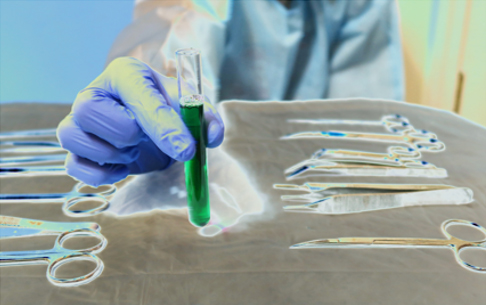 The water-soluble ICG-p28 solution can help surgeons to clearly define tumor margins and foci of cancer cells in
As described recently in Journal of Medicinal Chemistry, Dr. Yamada and his research team investigated the pharmacokinetics, biodistribution, and intraoperative imaging performance of ICG-p28 in breast cancer cell lines and animal models, with support from a Fiscal Year 2016 Breast Cancer Research Program Breakthrough Award - Funding Level 2. In animal models of two different types of breast cancer (estrogen receptor-positive and triple-negative breast cancer), rapid tissue uptake and localization at the tumors was observed following systemic injection of ICG-p28. Kinetic parameters and biodistribution analyses following ICG-p28 injection were comparable among all breast cancer cell lines, regardless of hormone receptor expression status, suggesting that ICG-p28 may potentially be suitable for a range of breast cancer types.
When tested as an intraoperative imaging agent using a transgenic animal model of breast cancer, the researchers found targeted delivery of ICG-p28 as well as a clear contrast of the mammary tumor relative to normal tissue. Moreover, the rate of tumor recurrence in animals, where ICG-p28 was used to guide surgical removal of tumors, was significantly decreased compared to the controls. Finally, when tested in numerous breast cancer cell lines, ICG-p28 did not cause any dose-related effects or significant toxicity.
These promising findings from Dr. Yamada's preclinical research support the potential development of ICG-p28 for use as an intraoperative imaging agent in patients undergoing BCS and may aid in the transition of this agent to early phase clinical trials. Ultimately, successful development of a tumor-targeted, intraoperative imaging agent for visual identification and complete removal of breast cancer cells during BCS has potential for significant impact on patient outcomes by reducing the chances for recurrence.
Publication:
Naffouje SA, Goto M, Coward LU, et al. 2022. Nontoxic tumor-targeting optical agents for intraoperative breast tumor imaging. Journal of Medicinal Chemistry 65(10):7371-7379. doi: 10.1021/acs.jmedchem.2c00417
Last updated Thursday, November 6, 2025