- Successful Isolation of BRCA2 (breast cancer type 2 susceptibility protein) and Elucidation of Its Mechanistic Functions Underlying Breast Cancer Tumorigenesis
- Effects of Aerobic Training on Cardiovascular Function and Tumor Biology in Women Receiving Neoadjuvant Chemotherapy for Operable Breast Cancer
- Breast Cancer Stem Cells: Multidrug Resistance and Survival in the Bone Marrow Niche
- Characterization of DCIS Progenitor Cells Reveals New Therapeutic Target
- Computational Modeling of Gene Network States and Drug Responses
- A Novel TGFβ Mouse Model of Breast Cancer Metastasis
- Microenvironmental Regulation of Mammary Carcinogenesis
- Priming Breast Tumors for Better Chemotherapeutic Response
- Development and Evaluation of a Novel Radiation Therapy Technique for the Treatment of Vertebrae with Metastatic Lesions
Successful Isolation of BRCA2 (breast cancer type 2 susceptibility protein) and Elucidation of Its Mechanistic Functions Underlying Breast Cancer Tumorigenesis
Posted October 27, 2010
Wolf-Dietrich Heyer, Ph.D. and Stephen C. Kowalczykowski, Ph.D., University of California, Davis, California
Deleterious mutations in the BRCA2 gene predispose individuals to hereditary breast, ovarian, and other cancers, and lead to startling increases in lifetime cancer risk. For example, BRCA2 mutation carriers have a 56%-87% increase in breast cancer risk by age 70 compared to an 8% risk in the general population.
The BRCA2 tumor suppressor gene was first discovered in 1994, but the purification of its full-length protein has taken nearly two decades due to its large size. BRCA2 is implicated in the regulation of Rad51-mediated homologous recombination, a process that repairs DNA breaks. Although it is well known that some mutations in BRCA2 cause cancer, the precise biological function of the BRCA2 protein remained largely unknown until August 2010, when three labs reported on the isolation of the full-length human BRCA2 protein and its role in DNA repair. Two of these three labs are led by Breast Cancer Research Program (BCRP) Idea Award recipients Drs. Wolf-Dietrich Heyer and Stephen Kowalczykowski, both from the University of California-Davis.
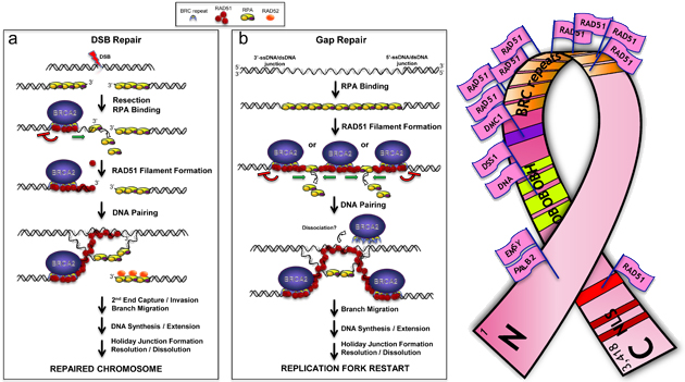
After receiving his award in 1999, Dr. Heyer quickly began the biochemical assessment of BRCA2. While various roles had been proposed for BRCA2, a direct interaction between BRCA2 and Rad51 was established, although the precise molecular activities of BRCA2 had not been identified. During the award period, Dr. Heyer began testing his hypothesis that BRCA2 protein is a critical accessory protein to Rad51 in the recombinatorial DNA repair pathway by purifying fragments of BRCA2 and assessing their interaction with Rad51 and DNA. Dr. Heyer and his colleagues recently reported that they successfully purified the full-length human BRCA2 protein after heterologous expression in yeast. Functional studies indicate that the primary mode of BRCA2 action is facilitating the assembly of Rad51 onto single-stranded DNA (ssDNA), which displaces replication protein A (RPA) to permit Rad51 to pair homologous DNA strand [1].
Using funds from his fiscal year 2008 award, Dr. Kowalczykowski purified the full-length human BRCA2 protein after overexpression in human cells and found that it stimulates Rad51-mediated DNA recombination by targeting Rad51 to ssDNA. Dr. Kowalczykowski found that BRCA2-mediated DNA repair is a consequence of several mutually reinforcing effects. Results indicate that BRCA2 enforces binding of Rad51 to ssDNA, accelerates the rate of RPA-displacement from ssDNA by Rad51 (a key regulatory step in DNA pairing), inhibits the ATPase activity of Rad51, and limits Rad51 binding to double-stranded DNA (dsDNA). Moreover, human BRCA2 is unable to anneal ssDNA-RPA complexes, the physiological intermediates of recombination, and this function may be assumed by Rad52, another protein involved in Rad51-mediated DNA recombination and repair [2].
Interestingly, the two scientists independently reached the same conclusion: BRCA2 assists Rad51-mediated homologous recombination of DNA by binding up to six RAD51 molecules per BRCA2 molecule. Mutations in BRCA2 can disrupt the DNA repair processes, leading to chromosomal instability and, eventually, cancer. The purification of full-length BRCA2 protein has opened doors for extensive studies on its function, including the mechanisms underlying BRCA2 deficiency-mediated oncogenesis. As Dr. Heyer states, "It took us a while to get this project developed, but it was the DoD funds that got this project started. This is an ideal example [of] how an Idea Award can initiate a breakthrough."
Publications:
1. Liu J, Doty T, Gibson B, and Heyer W-D. 2010. Human BRCA2 protein promotes RAD51 filament formation on RPA-covered single-stranded DNA. Nature Struct Molec Biol. Advance online publication doi:10.1038/nsmb.1904.
2. Jensen RB, Carreira A, and Kowalczykowski SC. 2010. Purified human BRCA2 stimulates RAD51-mediated recombination. Nature. Advance online publication doi:10.1038/nature09399.
Links:
Effects of Aerobic Training on Cardiovascular Function and Tumor Biology in Women Receiving Neoadjuvant Chemotherapy for Operable Breast Cancer
Posted October 22, 2010
Lee W. Jones, Ph.D., Duke University Medical Center, Durham, North Carolina
Anthracycline-containing chemotherapy improves disease-free survival and overall survival in women with early-stage or locally advanced breast cancer. However, use of anthracycline-containing chemotherapy is limited by two major problems: (1) poor solid tumor penetration as a result of inefficient tumor blood flow and hypoxia, and (2) dose-dependent, progressive cardiac dysfunction. Many strategies have been explored to independently address these issues with limited success.
Dr. Lee Jones, recipient of a Fiscal Year 2005 Breast Cancer Research Program Idea Award, has been exploring whether exercise training can improve drug delivery of chemotherapy and, simultaneously, protect the heart and cardiovascular system from the harmful effects of this therapy in women with locally advanced breast cancer. To address this question, Jones randomized 20 women receiving neoadjuvant chemotherapy (i.e. doxorubicin and cyclophosphamide) to usual care (i.e. chemotherapy alone) or chemotherapy plus supervised exercise training. Exercise training consisted of three moderate- to high-intensity cycle ergometry sessions per week for 12 weeks. At the beginning and end of the intervention, all participants completed assessments of exercise capacity, heart and vascular function, tumor blood flow, and provided a tumor biopsy. During the award period, Dr. Jones established that exercise training was safe and feasible for patients undergoing anthracycline-containing chemotherapy. Specifically, all women completed the study, and the attendance rate in the exercise group was 84 ± 5% (range, 0% - 100%), with women completing a mean of 30 exercise sessions out of 36 planned sessions. Preliminary results indicated that exercise capacity increased by 13% in the exercise group, and decreased by 10% in the chemotherapy-alone group. Remarkably, in healthy individuals, fitness typically declines ~10% every decade, suggesting that 12 weeks of chemotherapy causes a decade of physiological aging. In addition, there was evidence for cardiac injury in the chemotherapy-alone group that was prevented with exercise training. Dr. Jones is currently analyzing whether exercise improved chemotherapy delivery and efficacy in this study.
This study is the first to examine the effects of exercise training on cardiovascular function in women receiving neoadjuvant chemotherapy, and it is also the first to examine the effects on tumor biology in any cancer population. Dr. Jones believes that this work will provide new insights into the adverse cardiovascular effects of standard cancer chemotherapy and mechanisms by which exercise may prevent breast cancer recurrence.
Links:
Breast Cancer Stem Cells: Multidrug Resistance and Survival in the Bone Marrow Niche
Posted October 13, 2010
Peter V. Hauschka, Ph.D., Children's Hospital Boston, Boston, Massachusetts
Breast cancer recurrence may be due to the existence of cancer stem cells, which are theorized to arise from mutations in normal regenerative breast epithelial stem cells. Cancer stem cells not only initiate tumor growth, but also cause relapse by forming new tumors after treatment. Moreover, breast cancer preferentially metastasizes to bone, and the underlying mechanisms are not well characterized. Dr. Peter Hauschka, recipient of a Fiscal Year 2005 Breast Cancer Research Program Idea Award, hypothesizes that breast cancer initiation and therapeutic resistance are driven by cancer stem cells that have intrinsically high levels of drug efflux protein pumps (such as ABCG2/BCRP [breast cancer resistance protein] and MDR3 [multidrug resistance-3]) that actively expel chemotherapeutic molecules from the cells, giving them the ability to resist almost every therapeutic agent.
During the award period, research fellow Dr. Zhiyuan Zhang and Dr. Hauschka found that enhanced Notch3 expression may be a critical aspect of breast cancer bone metastasis. First, putative breast cancer stem cells were isolated from a population of the MDA-MET breast cancer cell line, which is known to preferentially metastasize to bone in animal models. When MDA-MET stem cells were grown in the presence of mouse osteoblasts, they resisted doxorubicin treatment, and exhibited increased colony formation. Additionally, the stem cells exhibited increased expression of mesenchymal markers (Notch3 and its ligand Jagged-1), i.e. genes that contribute to cancer invasiveness, which was stimulated by TGF-beta1 secreted by the osteoblasts. Inhibiting Notch3 expression after intracardiac injection of MDA-MET cells into mice significantly reduced bone marrow cancer colonization, while intact Notch3 expression allowed tumor cells to rapidly fill in the bone marrow space and form multiple osteolytic lesions in the animals.
Dr. Hauschka plans on pursuing whether Notch3 signaling is involved in breast cancer colonization to other metastatic sites, such as lung and brain. Interestingly, Notch3 signaling is generally correlated with poor clinical prognosis in Her2-negative breast cancer, which accounts for 70-80% of total incidence of primary and metastatic breast cancer, but not Her2-positive breast cancer. The Notch3 signaling pathway may be a novel molecular target for the treatment of Her2-negative breast cancer.
Publications:
Zhang Z, Wang H, Ikeda S, Fahey F, Bielenberg D, Smits P, Hauschka PV. Notch3 in human breast cancer cell lines regulates osteoblast-cancer cell interactions and osteolytic bone metastasis. Am J Pathol 2010 Sep;177(3):1459-69. Epub 2010 Jul 22.
Links:
Characterization of DCIS Progenitor Cells Reveals New Therapeutic Target
Posted July 8, 2010
Lance Liotta, George Mason University, Manassas, Virginia
Kirsten Edmiston, Inova Fairfax Hospital, Falls Church, Virginia
Approximately one-fourth of women diagnosed with breast cancer have ductal carcinoma in situ (DCIS), a nonaggressive, malignant precursor form of breast cancer that is confined to the mammary duct. Although most DCIS lesions remain dormant and do not invade or spread to the lymph nodes, some lesions progress to eventually become invasive and metastatic. Currently, there are no methods to predict which DCIS lesions will become invasive and no therapeutic options to prevent the invasive phenotype. Moreover, about 50% of high-grade DCIS lesions are estrogen receptor (ER)-negative, leaving these patients with limited options to treat their breast cancer.
Drs. Lance Liotta and Virginia Espina of George Mason University and Dr. Kirsten Edmiston of Inova Fairfax Hospital received a Synergistic Idea Award from the Fiscal Year 2006 Breast Cancer Research Program (BCRP). They hypothesized that some DCIS lesions are preprogrammed with invasive properties and that the mammary duct microenvironment provides a unique niche for DCIS cell survival. DCIS cells were isolated and propagated from xenotransplanted human DCIS lesions, and the cell cultures formed differentiated three-dimensional (3-D) ductal structures and adjacent stroma. DCIS cells demonstrated invasive properties by invading autologous stroma or xenografts. Protein array analysis revealed a set of activated signaling pathways consistent with a progenitor cell phenotype, including activated autophagy and mTOR/PI3K signaling pathways. Moreover, targeting the autophagy pathway with the drug chloroquine inhibited the propagation, invasion, and 3-D growth of the DCIS progenitor cells. These results suggest that autophagy may play a key role in regulating the emergence of DCIS invasive progenitor cells and that chloroquine is a potential new therapeutic for treating DCIS (Espina et al. 2010). As a result of their BCRP-supported research, Drs. Liotta and Edmiston have initiated a neoadjuvant clinical trial using chloroquine with or without tamoxifen as a potential DCIS treatment regimen to prevent progression to invasive breast cancer.
Publications:
Espina V, Mariani B, Gallager R, et al. 2010. Malignant precursor cells pre-exist in human breast DCIS and require autophagy for survival. PLoS ONE (5)4:10240-10255.
Abstracts:
Espina V, Gallagher RI, Mariani BD, et al. 2010. Pre-existence of invasive cells in breast ductal carcinoma in situ. American Society of Clinical Oncology. Chicago, Illinois. Oral presentation, Abstract #10508.
Espina V, Gallagher RI, Mariani BD, et al. 2010. DCIS neoadjuvant therapy: Targeting the autophagy pathway in malignant precursor cells. American Society of Clinical Oncology. Chicago, Illinois. Brd. 56F.
Espina V, Pastore L, Adamo L, et al. 2009. Human ductal carcinoma in situ contains malignant progenitor cells. Annual Meeting, American Association for Cancer Research, Denver, Colorado.
Liotta L and Espina V. 2009. Deciphering the dominant signaling pathways: The power of proteomics. Eighth International Congress on the Future of Breast Cancer, Maui, Hawaii.
Wulfkuhle JA, Espina V, Gallagher RI, et al. 2008. DCIS stem cells - Investigating the metastatic phenotype. US Department of Defense Breast Cancer Research Program Era of Hope Meeting, Baltimore, Maryland.
Patents:
Espina V and Liotta LA. Targeting the autophagy pathway to treat pre-malignant stages of breast cancer. US Patent Application No. 61/184,302.
Liotta LA and Espina V. Primary human DCIS cell lines: Treatment screening tool for inhibitors of invasive DCIS. US Patent Application No. 61/179,145, 61/222,253.
Espina V and Liotta LA. DCIS stem cells: A new paradigm for invasion and metastasis. US Patent Application No. 61/164,621.
Computational Modeling of Gene Network States and Drug Responses
Posted June 18, 2010
John Albeck, Ph.D., Harvard University, Boston, Massachusetts
Many of the genes that give rise to cancer when mutated have been identified, and new drugs have been designed to target them. However, the ramifications and downstream effects of targeting one gene within a network of genes are still relatively unknown. Many targeted therapeutics will block one gene's ability to cause cancer but will also affect downstream genes leading to unanticipated detrimental effects. Dr. John Albeck was awarded a Fiscal Year 2007 Breast Cancer Research Program Multidisciplinary Postdoctoral Award to develop a computational model of the PI3K gene network to predict gene responses to targeted therapeutics. This computational model could enable physicians to better identify which drugs will be most effective in treating breast cancer patients.
The Multidisciplinary Postdoctoral Award mechanism was designed to provide an opportunity for postdoctoral fellows to obtain training in two or more diverse disciplines. Dr. Albeck is using a multidisciplinary approach to tackle an important problem in breast cancer, while developing expertise in proteomics, translational oncology, cell biology, and computational biology under the mentorship of Drs. Joan Brugge, Gordon Mills, and Walter Fontana.
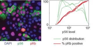
Dr. Albeck has developed a high-throughput immunofluorescence assay for signaling molecules in the PI3K/Akt/mTOR and several other related pathways, allowing for the ability to collect data on thousands of single cells per well across multiple 96-well plates, giving him a richer data set on cellular signaling states. Data from his studies have revealed a positive feedback loop between activated Akt and mTORC2 that may be the link in growth factor-mediated mTORC2 activation. Further studies will look into the factors that control cellular variability in the PI3K network, fluctuations in phosphorylation states, and the impact of pharmacological inhibitors.
Correlation of PI3K/Akt/mTOR activity and proliferative status a in heterogeneous population. Left: a high-throughput microscope image of DAPI (a marker for DNA present in all cells), pS6 (an indicator of mTOR activation) and pRb (an indicator of cell cycle commitment. Note the diversity of cellular states. Right: quantitative analysis of signaling states across the entire population reveals a strong positive correlation of cell cycle commitment over a very wide dynamic range of PI3K/Akt/mTOR signaling activity.
Links:
A Novel TGFβ Mouse Model of Breast Cancer Metastasis
Posted March 18, 2010
Manav Korpal and Yibin Kang, Ph.D, Princeton University, Princeton, New Jersey
More than 90 percent of breast cancer-related deaths are due to metastasis, a process that is dependent on interactions between tumor cells and the stroma of tissue at the metastasis site. To understand the signaling process that underlies breast cancer metastasis, Manav Korpal, recipient of a Fiscal Year 2007 Breast Cancer Research Program Predoctoral Traineeship Award, is focusing on the role of transforming growth factor beta (TGFβ) and the Smad pathway. His training with mentor Dr. Yibin Kang, a Fiscal Year 2005 BCRP Era of Hope Scholar Awardee, is fostering his development for a career as a breast cancer researcher, which is the ultimate goal of the Predoctoral Traineeship award mechanism.
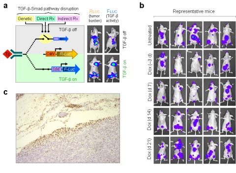
Using a novel mouse model in which metastasis and TGFβ signaling can be temporally and spatially monitored using non-invasive bioluminescence imaging technology, Mr. Korpal and Dr. Kang are examining TGFβ signaling in vivo in breast tumors with tropisms to bone, lung, and brain. The mouse model has enabled them to demonstrate the time-dependent efficacy of anti-TGFβ therapeutics on inhibiting the establishment of bone metastases. In addition, they were able to confirm the bone as a paracrine source of pathological TGFβ. This mouse model can also be used as an in vivo method to speed preclinical drug development through the evaluation of novel classes of TGFβ antagonists that are effective against breast cancer metastasis.
Early disruption of TGFβ signaling reduces establishment of bone metastases. a, A schematic illustration of the dual luciferase and Smad-inducible systems. The strength of the TGFβ pathway in tumor cells can be controlled by genetic manipulation of Smad4 expression, by direct pharmacological inhibitors of TGFβ receptor I kinases or through the use of indirect pharmacological inhibitors that reduce the bioavailability of TGFβ in the tumor microenvironment. FLUC, under the control of multiple SBEs, is expressed at low basal levels in the absence of TGFβ signaling (TGFβ off, top panel; upper right mouse image) relative to the TGFβ on condition (bottom panel; lower right mouse image). RLUC is expressed constitutively under the control of the CMV promoter, providing a measure for tumor burden (upper and lower left mouse images). b, Five representative mice from each group are shown in their supine position. Mice from the Dox (–3 d), Dox (d 7) and Dox (d 14) groups have reduced tumor burden in bone relative to untreated and Dox (d 21) mice suggesting that early treatment is more effective than late treatment. c, Gradient of p-Smad2 staining suggests that bone serves as a paracrine source of bioactive TGFβ.
Publications:
Korpal M, Yan J, Lu X, et al. 2009. Imaging transforming growth factor-beta signaling dynamics and therapeutic response in breast cancer bone metastasis. Nat Med 15:960-967.
Links:
Microenvironmental Regulation of Mammary Carcinogenesis
Posted March 10, 2010
Lisa Coussens, Ph.D., University of California, San Francisco, Helen Diller Family Comprehensive Cancer Center
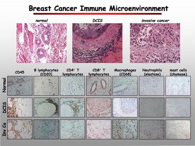
The human immune system serves a role in the prevention of cancer by identifying and destroying tumor cells; however, there is also evidence that the immune system may stimulate cancer growth and disease progression. Studies have shown that individuals suffering from chronic inflammation—wherein leukocytes, also called white blood cells, migrate incessantly into tissue—have an enhanced risk for cancer and that patients with malignant tissues containing leukocyte infiltrates tend to have a poor clinical prognosis compared to those without these infiltrates.
Dr. Lisa Coussens, a Fiscal Year 2006 Era of Hope Scholar awardee, is working to unravel the molecular mechanisms behind leukocyte infiltration into cancerous tissue and the subsequent leukocyte-mediated regulation of breast cancer progression. As part of her studies, Dr. Coussens is examining normal and malignant mammary tissues from both mice and humans to determine the types of immune cells present and to identify immune system factors that may mediate cancer. She has found that the infiltration of CD68+ macrophages, CD4+ and CD8+ T cells, and CD20+ B cells in human breast tumors parallels with advanced cancer progression and that immune expression signatures composed of macrophages and T cells could potentially be predictive for overall patient survival.
Dr. CoussensÂ’ in vivo studies have shown that infiltrating T cells promote metastasis to the lung in mice and provide evidence that the secretion of interleukin-4 (IL-4) by T cells enables cancer cells to migrate from the primary tumor site. Furthermore, she observed that a breast cancer model of mice deficient in leukocyte cysteine protease (cathepsin C) show a diminished number of lung metastases. Dr. Coussens is working to discern how T cells and cathepsin C promote lung metastasis and is examining the potential of cathepsin C expression as a predictive measure of metastasis.
If Dr. CoussensÂ’ work to delineate the molecular mechanisms behind leukocytemediated regulation of tumor cells is successful, she may potentially reveal molecules or pathways that could be therapeutic targets and may be used in the treatment of both primary tumors and metastatic disease.
Publications:
DeNardo DG, Barreto JB, Andreu P, Vasquez L, Tawfik D, Kolhatkar N, and Coussens LM. 2009. CD4+ T cells regulate pulmonary metastasis of mammary carcinomas by enhancing protumor properties of macrophages. Cancer Cell.
Links:
Public and Technical Abstracts: Microenvironmental Regulation of Mammary Carcinogenesis
Priming Breast Tumors for Better Chemotherapeutic Response
Posted February 8, 2010
Ralph Reisfeld, Ph.D., The Scripps Research Institute CaliforniaDr. Ralph Reisfeld received a Fiscal Year 2005 Breast Cancer Research Program Idea Award to investigate a new treatment option for breast cancer. Dr. Reisfeld hypothesized that the eradication of fibroblasts in the tumor stroma by a DNA vaccine that specifically targets the fibroblast activation protein (FAP) could increase uptake and sensitivity to chemotherapeutic agents. Dr. Reisfeld developed an oral vaccine that induces CD8+ T-cell-mediated immune responses against FAP. This vaccine suppressed tumor growth and experimental metastases in mice. When paired with doxorubicin or paclitaxel, the vaccine is more effective than single therapy with these therapeutic agents. Mice that received doxorubicin and vaccine treatment showed a decrease in metastatic infiltration in lung tissue. Dr. Reisfeld also discovered that the combination therapy resulted in increased apoptosis, or cell death, in tumors from these mice but had no detrimental effect on normal tissue. Furthermore, he found several proteins whose expression was reduced after treatment with vaccine and doxorubicin, for example, collagen type I expression, PDGF-C, VEGF-C, and VEGF-A, all known inducers of angiogenesis and lymphangiogenesis. Another beneficial effect of this combination therapy was the reduction of tumor interstitial fluid pressure, which correlates with increased tumor size. This combination therapy, specifically targeting fibroblasts, could bridge the gap between traditional chemotherapies and combinatory strategies for treating metastatic breast tumors.
Publications:
Liao D, Luo Y, Markowitz D, Xiang R, and Reisfeld RA. 2009. Cancer associated fibroblasts promote tumor growth and metastasis by modulating the tumor immune microenvironment in a 4T1 murine breast cancer model. PLoS One 4 (11):e7965.
Links:
Development and Evaluation of a Novel Radiation Therapy Technique for the Treatment of Vertebrae with Metastatic Lesions
Posted January 14, 2010
Joyce Keyak, Ph.D., University of California, Irvine
Breast cancer cells demonstrate strong predilection for metastasis to bone, especially the spine. Metastases to the spine of cancer patients can result in excruciating pain, vertebral collapse, and neurologic complications. One treatment option for patients with spinal metastases is vertebroplasty, a minimally invasive procedure in which a needle is inserted into the spine to extract the tumor tissue and replace it with bone cement. The cement then hardens to stabilize and restore the bone, often providing immediate pain relief. Usually, this surgical treatment is followed by multiple external beam radiation treatments to kill residual tumor tissue in the bone. Because irradiation of spinal metastases also exposes the spinal cord and nerves to radiation, the dose of radiation that can be delivered to the residual tumor is often limited.
Dr. Joyce Keyak, a Fiscal Year 2006 Synergistic Idea Award recipient, is working on combining radiation treatment with vertebroplasty to deliver more effective, therapeutic doses of radiation to patients afflicted with spinal metastases and to eliminate the need for numerous post-surgical clinical visits to receive radiation treatment. Working with cadaveric human spines, Dr. Keyak is studying the fabrication of radioactive bone cement, which would be injected following removal of cancerous tissue from the spine as a way to administer radiation therapy. Following identification of appropriate radioactive compounds for mixing with bone cement, Dr. Keyak performed studies that showed that the bone near the injected radioactive cement receives a high radiation dose and that the dose decreases rapidly with distance, so that the spinal cord will receive little, if any, radiation. Moving forward, Dr. Keyak plans to design and develop surgical instruments that can be used clinically for this procedure, to continue work on the development of quantitative treatment guidelines, and to apply to the FDA for approval to begin clinical trials. If successful, this work could have an important impact on the treatment of debilitating spinal metastases and improve the quality of life for patients receiving treatment for metastatic disease of the spine.
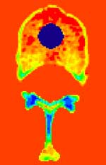
Part of a model for calculating radiation dose to a vertebra. Radioactive cement is shown as a dark blue circle; the bone is surrounded by a tissue equivalent (shown in red), and the other colors indicate various densities of bone (blue is the most dense and red is the least dense). (Graphic by Tadashi Kaneko, Ph.D., Keyak Laboratory, University of California, Irvine)
Links: