


- Targeting Detyrosinated Microtubule Protrusions to Reduce Breast Tumor Metastasis
- Combination Treatment of HER2/neu Expressing Breast Cancer Metastasis by Targeted alpha-Particle Radioimmunotherapy and Cancer Vacciner
- Nobel Prize in Chemistry Awarded to Breast Cancer Researcher
- Barriers to Breast Cancer Screening Among Latinas in the U.S.-Mexico Border Region
- Therapeutic Potential of Clostridium Perfringens Enterotoxin in Breast Cancer Brain Metastasis
- What a Concept! An Innovative Funding Mechanism for Breast Cancer Research
- Improving the Efficacy of Antiestrogen-Based Breast Cancer Therapies
- Breast Cancer Susceptibility Depends on the Genetic Ancestry of Latina Patients
- Tumor Stroma Predicts Clinical Outcome in Breast Cancer
- The Crystal Structure of a Potential Tumor Suppressor Bound to Its Target
- Genetic Counseling for Breast Cancer Susceptibility in African American Women
- Characterization and Restoration of Estrogen Sensitivity in Breast Cancer
- α6β4 Integrin Activation of NFκB-dependent Apoptosis Resistance in 3D Mammary Acini
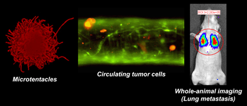
Targeting Detyrosinated Microtubule Protrusions to Reduce Breast Tumor Metastasis
Posted December 29, 2008
Stuart Martin, Ph.D., University of Maryland, Baltimore, Maryland

Metastatic disease is notoriously non-responsive to extant breast cancer therapy, and is the most common cause of death from breast cancer. Metastasis of breast cancer is a complex, multi-step event, and greater understanding of the mechanisms and characteristics of invasive, circulating tumor cells is crucial to developing therapies to combat metastasis.
Dr. Stuart Martin was awarded a fiscal year 2004 (FY04) Breast Cancer Research Program (BCRP) Concept Award to study apoptotic resistance of circulating mammary epithelial cells. During these studies he observed that these circulating cells produce long and dynamic protrusions that he initially believed to be actin-based invadopodia. Further examination revealed the protrusions were based on a specific, post-translationally modified form of α-tubulin, and that aggressively invasive breast cancer cell lines show higher frequencies of these protrusions than less invasive tumor cells. Dr. Martin named the protrusions "tubulin microtentacles," and he hypothesized that attachment to vascular walls via these protrusions might allow circulating tumor cells to avoid apoptosis or fragmentation in narrow capillaries. This hypothesis became the focus of the study proposed by Dr. Martin for his FY06 Idea Award.
Under this FY06 Idea Award, Dr. Martin identified that microtentacles result from the kinesin-dependent coordination of detyrosinated alpha-tubulin and vimentin intermediate filaments. In partnership with imaging experts, he has developed advanced methods to image microtentacle-bearing tumor cells circulating in intact blood vessels. Currently, Dr. Martin is refining and improving these imaging techniques in order to identify the molecular mechanisms underlying microtentacles, and define the steps undertaken by cells to anchor themselves to vascular vessel walls. Furthermore, he has identified several drug substances whose inhibitory effect on microtentacles may make them effective, targeted, anti-metastatic therapies. Together with clinicians at the University of Maryland Greenebaum NCI Cancer Center, Dr. Martin is isolating circulating tumor cells from breast cancer patients to test the prognostic and therapeutic potential of microtentacles. If successful, the application of neoadjuvant microtentacle-directed therapies could result in an urgently needed reduction in breast tumor metastasis.
Link:
Reducing the survival of circulating breast tumor cells (Adobe file) - Not able to view the embedded video in the file? You may need to download the free Microsoft Windows Media Player.
SELECT PUBLICATIONS:
Whipple RA, Balzer EM, Cho EH, et al. 2008. Vimentin filaments support extension of tubulin-based microtentacles in detached breast tumor cells. Cancer Res 68:5678-88.
Whipple RA, Cheung AM and Martin SS. 2007. Detyrosinated microtubule protrusions in suspended mammary epithelial cells promote reattachment. Exp Cell Res 313(7):1326-1336.
Combination Treatment of HER2/neu Expressing Breast Cancer Metastasis by Targeted alpha-Particle Radioimmunotherapy and Cancer Vacciner
Posted November 19, 2008
Hong Song, Ph.D., The Johns Hopkins University School of Medicine, Baltimore, Maryland
The Multidisciplinary Postdoctoral Award supports exceptionally talented postdoctoral fellows in obtaining significant training and experience in two disciplines so that they may more effectively pursue an independent career at the forefront of breast cancer research. As a recipient of a fiscal year 2004 Multidisciplinary Postdoctoral Award, Dr. Hong Song of Johns Hopkins University has received training in the areas of breast cancer radioimmunotherapy and biochemistry under the mentorship of Dr. George Sgouros and Dr. Todd Reilly. Dr. Song sought to combine targeted alpha-particle radioimmunotherapy and a whole-cell cancer vaccine to develop novel treatments for breast cancer. He studied the combined efficacy of 213Bi labeled anti-HER-2/neu antibody Fab' fragment and GM-CSF secreting 3T3 cells that express HER-2/neu in an immune-tolerant HER-2/neu transgenic mouse model. The combined treatment demonstrated improved efficacy in a mouse model of residual micrometastasis, compared to localized radiation or cancer vaccine alone, by reducing tumor growth and improving survival. The addition of the chemotherapy drug cyclophosphamide, which inhibits the T regulatory cell population, further improved efficacy of the combined treatment. These findings indicate that the combined treatment can specifically deliver radiation to HER-2/neu-positive tumor cells and boost the antitumor immune response, representing a new potential therapeutic approach. Dr. Song further used his multidisciplinary training to develop a preclinical model that can be used to evaluate immune-mediated metastatic breast cancer therapies. The new mouse model, which involves injection of the NT2.5 mammary tumor cell line into transgenic neu-N mice, enabled Dr. Song and colleagues to perform an array of imaging techniques to detect metastatic foci and potentially monitor response to immunotherapies.
Publication:
Song H, Shahverdi K, Huso DL, Esaias C, Fox J, Liedy A, Zhang Z, Reilly RT, Apostolidis C, Morgenstern A, and Sgouros G. 2008. 213Bi (alpha-emitter)-antibody targeting of breast cancer metastases in the neu-N transgenic mouse model. Cancer Research 68(10):3873-3880.
Song H, Shahverdi K, Huso DL, Wang Y, Fox JJ, Hobbs RF, Gimi B, Gabrielson KL, Pompser MG, Tsui BM, Bhujwalla Z, Reilly RT, and Sgouros G. 2008. An immunotolerant HER-2/neu transgenic mouse model of metastatic breast cancer. Clinical Cancer Research 14(19):6166-6124.
Nobel Prize in Chemistry Awarded to Breast Cancer Researcher
Posted October 29, 2008
Dr. Roger Tsien, University of California, San Diego, California

Dr. Roger Tsien, a Department of Defense Fiscal Year 2004 Breast Cancer Research Program (BCRP) Innovator Award grantee, has been selected by the Royal Swedish Academy of Sciences as a recipient of the 2008 Nobel Prize in Chemistry. Dr. Tsien is being recognized for his pioneering research in developing green fluorescent protein (GFP) and GFP derivatives that emit other colors into powerful molecular biology tools that are now routinely used by scientists in basic research on cancer and other diseases. His work is just one example of the way the Innovator Award fosters researchers of demonstrated creativity to shift their focus onto the enormous challenges of breast cancer.
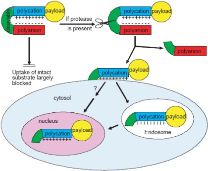
Applications of GFP or its derivatives require genetic modification of cells or organisms. Therefore, GFP or its derivatives are useful for tracking experimentally induced tumors in animals, but not cancers in human patients. Currently, Dr. Tsien is using his BCRP Innovator Award to develop a novel alternative mechanism to target tumor cells for imaging and drug delivery without requiring any gene transfer. This mechanism is based on "activatable cell-penetrating peptides" (ACPPs). If successful, ACPPs would be applicable to a wide range of imaging applications, including fluorescence, magnetic resonance imaging, gamma scintigraphy, and positron emission tomography, and to therapeutic agents such as chemotherapeutic drugs or radiation sensitizers. These techniques would allow for tailored imaging and therapeutic options based upon the specific needs of patients.
Certain arginine (Arg)-rich peptides known as "cell-penetrating peptides" can import covalently attached payloads into cells, but this uptake is not selective for cancer cells. Dr. Tsien's tumor-targeting strategy is to connect such a cell-penetrating peptide via a special linker to a short, negatively charged sequence. This composite molecule is an ACPP. The negative charges neutralize the positive charges, forming a hairpin structure that prevents cellular entry of the ACPP and attached payload. To target cancer cells, the linker region is designed to be specifically cleaved by extracellular proteases such as matrix metalloproteinases (MMPs), which are concentrated in the tumor microenvironment. Normal cells cannot cut the linker, so they leave the hairpin intact and do not take up the ACPP. However, the proteases in the tumor microenvironment cleave the linker, releasing the Arg-rich peptide sequence and the associated drug or contrast agent to adhere to and be taken up by tumor cells in the immediate vicinity.
Current ACPPs enable tumors to be seen both by magnetic resonance imaging (MRI), for whole-body scanning, and by far-red fluorescence, for real-time guidance during surgery. This combination in a single ACPP enables preoperative delineation of tumors by MRI, more accurate and complete removal by fluorescence-guided surgery, and postoperative review by MRI. These advances are improving long-term survival in two syngeneic tumor models. ACPPs can also image primary tumors and lung metastases in the transgenic MMTV-PyMT breast cancer mouse model, as well as malignant regions in resected human breast cancer tissue samples. Additionally, Dr. Tsien's group has developed ACPPs activated by enzymes other than MMPs, thus broadening the range of tumors and diseases to which this technology is applicable. He is currently trying to maximize the contrast between tumors and normal tissues, to make ACPPs synergistic with other established targeting mechanisms such as antibodies, to improve delivery of chemotherapeutic agents, and to advance these new molecules into clinical trials. With a potentially wide range of imaging and therapeutic options amenable to ACPP-mediated delivery, the research supported by Dr. Tsien's BCRP Innovator Award may lead to new and powerful treatment modalities that combine imaging with therapy to facilitate personalized treatment of breast cancer.
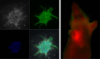
The left panels show 3D images of protease activatable peptides (ACPPs) being taken up into invading mammary adenocarcinoma cells grown in 3D culture (peptide fluorescence in white, counterstain for live cells in green, counterstain for cell nuclei in blue, and overlaid composite at lower right). The right panel shows fluorescence of doxorubicin-loaded nanoparticles (red) used as therapeutic components for ACPPs, accumulating in a xenografted tumor in a mouse. Green denotes autofluorescence.
Barriers to Breast Cancer Screening Among Latinas in the U.S.-Mexico Border Region
Posted October 23, 2008
Jose A. Pagan, Ph.D., The University of Texas-Pan American, Edinburgh, Texas

Dr. Jose Pagan, the Principal Investigator of a Fiscal Year 2005 Historically Black Colleges and Minority Institutions Partnership Training Award, is a faculty member at the Institute for Population Health Policy (IPHP) at the University of Texas-Pan American (UTPA). Under the HBCU/MI Partnership Training funding mechanism, Dr. Pagan and his colleagues participate in a mentoring partnership with established breast cancer researchers at the Leonard Davis Institute of Health Economics at the University of Pennsylvania. Through this partnership, the IPHP can leverage its existing expertise in conducting health and social/behavioral research-particularly as it relates to low-income Latino populations-to develop a long-term, successful, and competitive breast cancer research program at UTPA.
In 2008, Dr. Pagan published results of a study he performed on data collected from the 2000-2001 Community Tracking Study Household Survey (CTSHS). The CTSHS is a national survey dataset from households throughout the United States that tracks health care coverage and use, in addition to other data. Dr. Pagan showed that uninsured women were less likely to have undergone mammography than insured women. Furthermore, the study showed that in local populations where the uninsured rate was high, all women, whether they had insurance or not, were less likely to get a mammogram.
Currently, Dr. Pagan is conducting a survey of nearly 900 Latinas in the South Texas region to determine community characteristics (e.g., marital status, education, time since immigration) that might have a connection to mammographic screening rates. He hopes to identify barriers to breast cancer screening in this population and help develop strategies to improve screening rate differences. Breast cancer is the most frequently diagnosed cancer in Latinas; achieving appropriate breast cancer screening levels has the potential to significantly affect the morbidity and mortality rates of breast cancer in this population.
Publication:
Pagan JA, Asch DA, Brown CJ, et al. 2008. Lack of community insurance and mammography screening rates among insured and uninsured women. Journal of Clinical Oncology 26(11):1865-1870.
Link:
Abstract: Barriers to Breast Cancer Screening Among Latinas in the U.S.-Mexico Border Region
Therapeutic Potential of Clostridium Perfringens Enterotoxin in Breast Cancer Brain Metastasis
Posted October 22, 2008
Scott L. Kominsky, Ph.D., Johns Hopkins University School of Medicine
While there have been marked improvements in the management of systemic breast cancer, metastasis to the brain still poses a significant challenge. The increased survival of breast cancer patients appears to be accompanied by an increased incidence of brain metastasis, likely due to the inability of systemic treatments to cross the blood brain barrier. Patients with brain metastases frequently die from neurological complications, and with standard treatment the average survival time is only about eight months. Furthermore, long-term survivors often suffer damaging side effects from their treatment. The diagnosis of breast cancer metastasis to the brain is only made following the development of symptoms. Therefore, the development of new and safer treatment options for brain metastases is of immediate clinical importance.
Dr. Scott Kominsky, who is supported by a Department of Defense Fiscal Year 2005 Breast Cancer Research Program Idea Award, has found that brain metastases express cell surface receptors for a cytotoxic enterotoxin that is produced by the bacterium Clostridium perfringens. These receptors, termed Claudin-3 and Claudin-4, are present only in metastatic cells in the brain, not in normal brain tissue. These receptors may therefore present a unique opportunity to locally treat and eliminate tumors without damaging the normal surrounding tissue. Dr. Kominsky has proposed to characterize Claudin-3 and 4 as specific markers of breast cancer brain metastases and to examine the ability of the C. perfringens enterotoxin (CPE) to eliminate these metastases without harming host brain tissue. By using state-of-the-art bioluminescent imaging technology, Dr. Kominsky is developing the first mouse model of breast cancer metastasis to the brain that permits the measurement of both tumor growth and response to treatment in live animals and in real time. To destroy metastatic cells in the brain, Dr. Kominsky and colleagues are incorporating CPE into biodegradable polymer micro-spheres that will be administered intracranially to tumors in their animal model. This approach circumvents the blood brain barrier and allows effective drug concentrations to be achieved at the tumor site while maintaining low systemic drug levels.
This work aims to provide a new therapeutic target for the treatment of breast cancer metastasis to the brain and a new animal model of breast cancer brain metastasis. Further, the potential of CPE to eliminate or significantly reduce tumor size without harming host brain tissue could extend patient survival and improve quality of life. In addition to targeting breast cancer metastases to the brain, this concept may also apply to other cancers whose epithelial brain metastases express Claudin-3 and -4.
Publication:
Kominsky SL, Tyler B, Sosnowski J, Brady K, Doucet M, Nell D, Smedley JG 3rd, McClane B, Brem H, and Sukumar S. 2007. Clostridium perfringens Enterotoxin as a Novel-Targeted Therapeutic for Brain Metastasis. Cancer Research 67:7977-7982.
Link:
What a Concept! An Innovative Funding Mechanism for Breast Cancer Research
Posted October 22, 2008
Jianghong Rao, Ph.D., Stanford University, Stanford, CA
Ruth Lupu, Ph.D., Evanston Northwestern Healthcare Research Institute, Evanston, IL
Albert J. Millis, Ph.D., State University of New York Albany, Albany, NY
The Breast Cancer Research Program (BCRP) Concept Award mechanism provides researchers of all levels the opportunity to explore innovative ideas that could open new doors in breast cancer research. This award mechanism is unique in that it does not allow preliminary data in the research proposal. Concept Awards fund newly conceptualized ideas that most likely would not have been supported through other funding sources. This award mechanism serves as a way for investigators to test new hypotheses, explore intriguing observations, and collect preliminary data that may be used in applying for future funding. For example, Drs. Jianghong Rao, Ruth Lupu, and Albert J. Millis each received a Fiscal Year 2005 Concept Award and have generated exciting results that could lead to new areas of breast cancer investigation.
Dr. Jianghong Rao has developed a novel nanosensing system designed to both detect and image matrix metalloproteinases (MMPs). MMPs are classified as a family of secreted endopeptidases, including collagenases, stromelysins, gelatinases, and membrane-type MMPs, that are involved in the regulated degradation and processing of extracellular matrices. These enzymes play a critical role in defining the cellular environment and are also upregulated in virtually all human cancers. MMPs have been associated with initiation, invasion, and metastasis of breast cancer. Dr. Rao has developed highly sensitive bioluminescent nanosensors to detect MMPs using Quantum dot (QD) technology (fluorescent semiconductor nanocrystals with surface carboxylic acids) combined with bioluminescent proteins. These nanosensors are made through a specific bioconjugation reaction of a fusion protein containing the MMP-2 substrate and the bioluminescence resonance energy transfer (BRET) donor, a mutant form of Renilla luciferase, to the QDs. In the absence of MMP-2, BRET occurs due to the close proximity between the QDs and the luciferase and light emission is produced from the QD. However, the cleavage of an amide bond by MMP-2 releases the luciferase protein and prevents BRET from occurring. With this system, Dr. Rao was able to detect 2 ng/ml of MMP-2, a sensitivity that improves upon that of fluorescence-based nanosensors. Dr. Rao demonstrated detection of MMP-2, MMP-7, and urokinase in buffer, tumor lysates, and mouse serum with the system, as well as simultaneous multiplex detection of MMP-2 and urokinase. This novel technology presents the possibility of real-time detection of the activity of MMPs in the blood, not only to further study the roles of this class of enzymes, but also to allow for the development of cancer diagnostic and drug screening assays. The results from this Concept award have allowed him to secure R01 funding from the National Cancer Institute to further pursue these goals.
Dr. Ruth Lupu has been investigating Cyr61, an angiogenic factor expressed in approximately 30 percent of estrogen receptor-negative invasive breast carcinomas. Cyr61 interacts with αvβ3, an integrin whose expression appears to correlate with a poor prognosis. Cyr61/αvβ3 interactions are believed to be critical in breast cancer progression, wherein the overexpression of Cyr61 and signaling through αvβ3 could lead to the development of a chemoresistant phenotype. In this project, Dr. Lupu tested antagonistic αvβ3 compounds in a dose-dependent study of breast cancer cells that overexpress Cyr61 and are resistant to Taxol cytotoxicity. As the dose of antagonist compounds increased, the efficacy of Taxol significantly increased in both a dose-dependent and αvβ3-specific manner. These results indicate that Cyr61/αvβ3 interactions cooperatively regulate breast cancer cell sensitivity to Taxol. Experiments to knock down Cyr61 expression using siRNA suggest that there is a critical balance between Cyr61 and αvβ3 expression. The findings generated from this project could potentially lead to the use of Cyr61 as a predictive marker for response to chemotherapies in breast cancer patients.
Dr. Albert J. Millis has investigated the use of high-affinity RNA aptamers that can facilitate reduction in the levels of proteins associated with the development and progression of breast cancer. These high affinity aptamers have the bi-functional capacity to bind and form bridges between the targeted protein and a cellular process that would facilitate its degradation. Specifically, targeted extracellular proteins would undergo opsonization and uptake via the iC3b receptor, and the internalized proteins would be targeted for degradation. Dr. Millis constructed an iC3b -green fluorescent protein (GFP) bi-functional aptamer, and tested for iC3b receptor-mediated uptake into the lysosomal compartment of human macrophages. The results of this study demonstrate a proof of concept that extracellular proteins can be targeted for cellular uptake via the complement system resulting in target protein degradation in lysosomes. This technology has broad applicability and could be adapted to validate drug targets, as well as to treat breast cancer patients by targeting specific proteins for their inactivation and/or destruction. High affinity bi-functional aptamers, with their ability to target specific proteins, could therefore be used to block tumor cell progression and metastasis at multiple stages of the disease.
Through the Concept Award mechanism and the research efforts of investigators like Drs. Rao, Lupu, and Millis, the BCRP has funded many highly innovative projects that have successfully tested a hypothesis and led to new avenues of investigation. The ability for investigators to explore novel ideas that are innovative yet risky represents a unique opportunity to make advancements in breast cancer research.
Publications:
Yao H, Zhang Y, Xiao F, et al. 2007. Quantum dot/bioluminescence resonance energy transfer based highly sensitive detection of proteases. Angewandte Chemie International ed. in English 46(23):4346-4349.
Xing Y, So M, Koh AL, et al. 2008. Improved QD-BRET conjugates for detection and imaging. Biochemical and Biophysical Research Communications 372:388-394.
Vellon L, Menendez J, Hong L, et al. 2007. Up-regulation of αvβ3 integrin expression is a novel molecular response to chemotherapy-induced cell damage in a heregulin-dependent manner. Differentiation 75(9):819-830.
Link:
Abstract: Interaction between Cyr61 and avbeta3 in Breast Cancer: Role in Taxane Resistance
Improving the Efficacy of Antiestrogen-Based Breast Cancer Therapies
Posted August 22, 2008
Johanna K. Perry, Ph.D., Liggins Institute, University of Auckland, Auckland, New Zealand
A majority of breast cancer cases in postmenopausal women are estrogen receptor (ER)-positive. Drugs that inhibit ER signaling, such as tamoxifen, are effective systemic treatments for these patients. For reasons that are unclear, however, a significant proportion of women will become resistant to these therapies due to the recurrence of endocrine-resistant breast cancer. Elucidation of the molecular mechanisms that underlie the development of this resistance could potentially lead to new drug targets for combinatorial therapies with tamoxifen. Recent reports suggest that expression of growth factors and/or their receptors by tumor cells is involved in resistance to anti-estrogen therapy, as this might bypass the need for ER-mediated growth stimulation. Indeed, autocrine expression of human growth hormone (hGH) has recently been implicated in the progression of human breast cancer.
Dr. Johanna Perry, who was supported by a Department of Defense Fiscal Year 2005 Breast Cancer Research Program Concept Award, investigated whether autocrine hGH could confer resistance to antiestrogen-based breast cancer therapies. Dr. Perry tested this hypothesis in a cell culture model in which either control human breast cancer cells or human breast cancer cells that overexpressed hGH would be incubated with the antiestrogen drugs tamoxifen and fulvestrant. In apoptosis and soft agar colony formation assays, autocrine hGH increased the resistance of these cells to drug-mediated cytotoxicity. Autocrine hGH also upregulated the mRNA levels, protein levels, and transcriptional activity of the estrogen biosynthesis enzyme aromatase. Cells that overexpressed hGH were more resistant to the cytotoxic effects of the aromatase inhibitor exemestane (Aromasin). Autocrine hGH-mediated upregulation of aromatase activity was found to be dependent on the kinase JAK-2, providing a starting point to further delineate this signal transduction pathway.
These results suggest that autocrine hGH may be a therapeutic target, and that functional antagonism of hGH-mediated signal transduction, either alone or as an adjuvant therapy, has potential therapeutic relevance for the treatment of ER-positive human breast cancer. As an FDA approved hGH receptor antagonist is already on the market, this project may result in rapid clinical application to improve the prognosis of patients with hormone sensitive breast cancer.
Link:
Abstract: Improving the Efficacy of Antiestrogen-Based Breast Cancer Therapies
Tumor Stroma Predicts Clinical Outcome in Breast Cancer
Posted July 16, 2008
Francois Pepin, McGill University, Montreal, Quebec, Canada
The pathology of breast tumors is diverse and sometimes deceiving. Breast cancer patients at the same cancer stage or whose tumors have similar pathology upon examination may respond differently when given the same treatment. Many researchers are working in pursuit of a better way to profile a tumor and predict clinical outcome to ensure the patient receives the best targeted treatment. Francois Pepin of McGill University was awarded a Fiscal Year 2005 Predoctoral Traineeship Award to apply genomic approaches to characterize the role of tumor stroma in breast cancer, under the mentorship of Michael Hallett and in collaboration with Morag Park. As a result of the collaboration between the Hallett and Park labs, Mr. Pepin has received strong multidisciplinary training in breast tumor biology, genomics, and bioinformatics. This collaborative, multidisciplinary group focuses on the tumor stroma, a lining of cells that surrounds the tumor and provides an essential role in tumor development by delivering nutrients through the supporting vasculature. Using tissue samples from patients with invasive breast cancer, laser capture microdissection was performed to isolate and compare stromal cells from normal mammary tissue and breast tumors. These samples were characterized using genetic microarray profiling and defined gene expression signatures based on variation between tumor tissue and normal stroma. On the basis of differential gene expression, the group has developed a 26-gene predictor called the stroma-derived prognostic predictor (SDPP) that can forecast disease outcome with greater accuracy than standard clinical prognostic factors and current predictors that are derived from whole tissue. The SDPP is capable of identifying individuals from several clinical subtypes who will have a poor disease outcome and predicting the type of treatment that may provide the greatest benefit to the individual. The SDPP could also help identify patients who may benefit from immunotherapy and distinguish between those who may or may not require aggressive treatment. The group also found that when the SDPP is used in combination with existing outcome predictors, the prognostic power of the SDPP increased substantially. The ability of the SDPP to differentiate tumor response to treatments emphasizes the importance of stromal biology in breast cancer diagnosis and the profiling of individual breast tumors for prediction of clinical outcome.
Selected Publication:
Finak G, Bertos N, Pepin F, Sadekova S, Souleimanova M, Zhao H, Omeroglu G, Meterissian S, Omeroglu A, Hallett M, and Park M. 2008. Stromal gene expression predicts clinical outcome in breast cancer. Nature Medicine 14(5):518-527.
Link:
Abstract: Genome-Wide Approaches To Detecting Stromal-Epithelial Interactions In Breast Cancer
Breast Cancer Susceptibility Depends on the Genetic Ancestry of Latina Patients
Posted July 8, 2008
Elad Ziv, M.D., University of California, San Francisco, California
Breast cancer incidence and mortality vary substantially among different racial and ethnic groups in the United States. The incidence of breast cancer is highest in Caucasian women, followed by African American, Asian, Latina, and Native American women. The role of genetic factors in explaining differences in incidence rates between racial/ethnic populations remains largely unexplored. The Latino population within the U.S. is an admixed group comprised of individuals with differing amounts of European, African, and Native American ancestries. Since these individual ancestries span the range from highest to lowest incidence of breast cancer in their respective homogeneous populations, the Latina group may therefore provide valuable insight into the genetic and environmental factors that contribute to disease susceptibility.
Dr. Elad Ziv, recipient of a Department of Defense Fiscal Year 2003 Breast Cancer Research Program Idea-Epidemiology Award, hypothesized that European ancestry among Latinas would be associated with a higher risk of breast cancer, and that inaccurate predictions of breast cancer risk in Latinas could be adjusted by determining their individual ancestries. Dr. Ziv proposed to correlate disease risk with individual genetic ancestry by using DNA tests to measure the relative numbers of DNA sequences, called ancestry informative markers, which can be used to statistically estimate the maximum likelihood of ancestry (i.e., European, African, etc.) in any individual. Consistent with the epidemiological data, this work identified in Latina women an association between higher European ancestry and increased breast cancer risk. Furthermore, Dr. Ziv and colleagues developed a novel algorithm that uses DNA sequences called single nucleotide polymorphisms to infer geographic ancestry solely from genotypic data.
These results suggest genes that are involved in breast cancer susceptibility may be identified by studying admixed populations. The identification of such genes may dramatically affect the practices of early breast cancer detection and prevention. Instead of uniform screening of all women beginning at a particular age, individual risk profiles based in part on genetic assessment of risk could lead to tailored screening recommendations, and could identify women who would particularly benefit from modification of disease-associated lifestyle factors.
Selected Publications:
Ziv E, John E, Kho J, et al. 2006. Breast cancer risk factors and genetic ancestry among Latinas in the San Francisco Bay area. Cancer Epidemiology Biomarkers and Prevention 15:1878-1885.
Paschou P, Ziv E, Burchard E, et al. 2007. PCA-correlated SNPs for substructure identification. PLOS Genetics 3:1672-1686.
Link:
Abstract: Admixture And Breast Cancer Risk Among Latinas
The Crystal Structure of a Potential Tumor Suppressor Bound to Its Target
Posted June 23, 2008
Mark A. Lemmon, Ph.D., University of Pennsylvania School of Medicine, Philadelphia, Pennsylvania
Cancer often results from the aberrant expression of signaling molecules that control cellular growth and differentiation. The four members of the ErbB family of receptor tyrosine kinases, which are activated by epidermal growth factor (EGF) and other ligands, have been strongly linked to breast cancer. ErbB2 is overexpressed in approximately 30% of breast cancer cases, and its targeting by the therapeutic antibody Herceptin improves the survival of these patients. However, recent clinical trials targeting other members of the ErbB receptor family have been disappointing. This may be due to ligand-dependent heterodimerization of the receptors for activation, which would also enable this pathway to remain active even when one of the receptors is blocked pharmacologically. In addition, at least 50% of breast cancer cases involve the expression of ErbB receptor ligands. Thus, inactivating or neutralizing the growth factor ligands that activate ErbB receptors may be an alternative therapeutic method for targeting ErbB signaling in breast cancer.
Dr. Mark Lemmon, supported by a Department of Defense Fiscal Year 2004 Breast Cancer Research Program Idea Award, proposed to target the ligands that activate ErbB receptors using a protein that acts as an antagonist of EGF receptor signaling in the fruit fly Drosophila melanogaster. Dr. Lemmon's group has found that the secreted protein Argos inhibits EGF receptor signaling by binding and neutralizing the native EGF-like ligand Spitz rather than the EGF receptor. Argos may therefore provide a model for the therapeutic blockade of ErbB signaling by ligand sequestration in humans. In a recent paper published in the journal Nature, Dr. Lemmon's group determined the crystal structure of a 1:1 complex of Argos and Spitz with a resolution of 1.6 ?. Argos contains a set of three domains that wrap around Spitz, similar to the way a C-clamp is fastened to a surface.
The solution of the crystal structure of Argos:Spitz may provide a new way to identify functional homologues of Argos in humans that may be exploited to inhibit EGF signaling in breast cancer. While Argos shares neither sequence nor structural homology with the EGF receptor, the researchers found unexpectedly that Argos shares structural homology with the ligand-binding domains of the human receptors for transforming growth factor-? and the urokinase-type plasminogen activator. Similarly, several human proteins of unknown function, including some that are implicated in metastatic cancer, are similar to Argos in structure, but not sequence. This study provides the starting point for rational design of novel proteins with higher affinity for EGF, potentially providing new avenues in the development of specific and potent breast cancer therapies.
Selected Publication:
Klein DE, Stayrook SE, Shi F, Narayan K, and Lemmon MA. 2008. Structural basis for EGFR ligand sequestration by Argos. Nature May 25 (Epub ahead of print).
Link:
Abstract: Harnessing Novel Secreted Inhibitors Of EGF Receptor Signaling for Breast Cancer Treatment
Genetic Counseling for Breast Cancer Susceptibility in African American Women
Posted March 27, 2008
Chanita Hughes, Ph.D., University of Pennsylvania, Philadelphia, Pennsylvania
Each year, thousands of African American women are diagnosed with breast cancer. Epidemiologic studies have shown that about 16% to 28% of African American women who have a personal and family history of breast and/or ovarian cancer carry mutations in the susceptibility genes BRCA1 and BRCA2 (BRCA1/2). Genetic counseling and testing for BRCA1/2 mutations is now available through clinical research programs using standard genetic counseling (SGC) protocols. However, African American and Caucasian women differ in their attitudes about and responses to education and counseling. Increasingly, cultural beliefs and values are being recognized as important factors in genetic counseling. Yet, despite recommendations to increase the cultural sensitivity of breast cancer risk counseling, such programs have not been developed or evaluated.
Dr. Chanita Hughes Halbert, sponsored by two Fiscal Year 1999 Breast Cancer Research Program awards - the Career Development Award and an Idea Award - proposed to develop a Culturally Tailored Genetic Counseling (CTGC) protocol for African American women and determine the protocol's impact on decision-making processes with regard to genetic testing for BRCA1/2 mutations and cancer prevention practices. The SGC and CTGC protocols both provide education about hereditary cancer, genetic testing, and risk information. However, consistent with guidelines for providing culturally competent genetic counseling, the CTGC protocol includes standardized probes to elicit discussion about cultural factors that have been shown to influence decisions about genetic counseling among African American women (e.g., spiritual and religious beliefs, communalism). Dr. Halbert's study randomly assigned African American women with a family history of breast and/or ovarian cancer to either the CTGC (n=87) or SGC group (n=94). A number of findings from this study have important implications in breast cancer in African American women. Most of the women in the study demonstrated a willingness to participate in genetic counseling research and had a high perception of the benefits of genetic testing. The study had high satisfaction (96%) and retention (60%) rates. However, only 30% of the participants agreed to undergo genetic testing. Dr. Halbert's research also found that the environmental setting from which the women were recruited (oncology facility vs. general medical practices), as well as the family history of breast/ovarian cancer, had a significant impact on the outcome of the genetic counseling. CTGC showed a clear advantage over SGC as measured by lessened worries and improved coping strategies. This suggests that there may be benefits to discussing cultural beliefs and values during genetic counseling with African American women. In conclusion, the culturally tailored protocol could be adopted as a new resource for genetic counselors to discuss breast cancer risk and prevention with African American women and other minority or ethnic populations.
Selected Publications:
Brewster K, Wileyto EP, Kessler L, Collier A, Weathers B, Stopfer JE, Domchek S, and Halbert CH. 2007. Sociocultural predictors of breast cancer risk perceptions in African American breast cancer survivors. Cancer Epidemiology, Biomarkers & Prevention 16:244-248.
Halbert CH, Kessler L, Stopfer JE, Domchek S, and Wileyto EP. 2006. Low rates of acceptance of BRCA1 and BRCA2 test results among African American women at increased risk for hereditary breast-ovarian cancer. Genetics in Medicine 8:576-582.
Halbert CH, Brewster K, Collier A, Smith C, Kessler L, Weathers B, Stopfer JE, Domchek S, and Wileyto EP. 2005. Recruiting African American women to participate in hereditary breast cancer research. Journal of Clinical Oncology 23:7967-7973.
Halbert CH, Kessler L, Collier A, Wileyto EP, Brewster K, and Weathers B. 2005. Psychological functioning in African American women at increased risk for hereditary breast and ovarian cancer. Clinical Genetics 68:222-227.
Halbert CH, Kessler LJ, and Mitchell E. 2005. Genetic testing for inherited breast cancer risk in African Americans. Cancer Investigation 23:285-295.
Kessler L, Collier A, Brewster K, Smith C, Weathers B, Wileyto EP, and Halbert CH. 2005. Attitudes about genetic testing and testing intentions in African American women at increased risk for hereditary breast cancer. Genetics in Medicine 7:230-238.
Characterization and Restoration of Estrogen Sensitivity in Breast Cancer
Posted March 4, 2008
Pamela Munster, Ph.D., H. Lee Moffitt Cancer Center & Research Institute, University of South Florida, Tampa, Florida
Efforts to combat estrogen receptor (ER)-positive breast cancers primarily involve inhibition of the estrogen receptor or targeting members of the ER pathway. ER-positive cancers are shown to respond effectively to selective estrogen receptor modulators (SERMs), selective estrogen receptor down-regulators (SERDs), and aromatase inhibitors. However, a number of patients do not respond or eventually develop resistance to anti-hormonal treatment. In vitro studies have shown that combining histone deacetylase (HDAC) inhibitors with anti-hormonal therapies can restore response to therapy, although the exact mechanisms underlying this synergy are not known. Dr. Pamela Munster, recipient of a Department of Defense Fiscal Year 2005 Breast Cancer Research Program Concept Award, is working to identify the specific HDAC enzymes that play a role in resistance to anti-hormonal therapy in ER-positive breast cancer.
The synergistic effects of HDAC inhibitors and the anti-estrogen tamoxifen were examined in breast cancer cells by selectively targeting HDAC1, 2, and 6 for deletion by siRNA. Western blot analyses demonstrated that a concomitant decrease in ER and progesterone receptor (PR) expression occurred with depletion of HDAC2 in breast cancer cell lines. In contrast, depletion of HDAC1 and HDAC6 decreased ER but not PR expression. Analyses by growth and apoptosis assays indicate that depletion of HDAC2, but not HDAC1 or HDAC6, enhanced breast cancer cell sensitization to tamoxifen. Additional studies showed that ER-positive and ER-negative breast cancer cells exposed to HDAC inhibitors such as valproic acid, hydroxamic acid, and benzamides resulted in decreased PR expression. Furthermore, cells exposed to a combination of tamoxifen and valproic acid exhibited enhanced downregulation of PR, suggesting a synergistic interaction between the HDAC inhibitors and tamoxifen. These results suggest that the development of HDAC inhibitors (specifically those that target HDAC2) in combination with SERMs may be a promising strategy for the treatment of breast cancer.
Currently, Dr. Munster is evaluating the expression of ER, PR, and HDAC as they correlate to patient response in conjunction with a Phase II trial of the HDAC inhibitor suberoylanilide hydroxamic acid (SAHA, Vorinostat) combined with tamoxifen for patients with advanced breast cancer who have failed prior anti-hormonal therapy. These studies will help to determine whether PR and HDAC levels may be used as predictors of response in ER-positive breast cancer patients.
Link:
Abstract: Characterization and Restoration of Estrogen Sensitivity in Breast Cancer
α6β4 Integrin Activation of NFκB-dependent Apoptosis Resistance in 3D Mammary Acini
Posted January 25, 2008
Valerie Weaver, Ph.D., University of California, San Francisco, California
Relatively little is known about how the three-dimensional (3D) tissue microenvironment of mammary tumors impacts cell survival and apoptosis. Defects in the regulation of apoptosis are thought to reduce the effectiveness of certain breast cancer treatments. Fiscal Year 2004 Era of Hope Scholar Dr. Valerie Weaver is studying the roles of integrins and the microenvironment in mammary epithelial cell (MEC) tissue architecture, cell survival, and regulation of the breast tumor phenotype.
Dr. Weaver and colleagues previously established a 3D model culture system with MECs that is designed to represent the cell-cell and cell-extracellular matrix interactions found in the breast stromal environment. Highlighting the importance of the cellular environment, they found that MECs in 3D culture form acini structures and show enhanced survival and increased resistance to cell death from various insults when compared to cells grown in 2D cultures. The apoptosis-resistant 3D mammary acini phenotype was mediated by α6β4 integrin-dependent tissue polarity through activation of the NFκB activation pathway in the absence of hypoxia and without regard for cell growth status. In recent studies published by Dr. Weaver's group, two non-malignant immortalized MEC lines were used to analyze the apoptotic resistant phenotype on laminin-rich basement membrane. They found that resistance to apoptosis occurred through increased levels of α6β4 integrin, thus initiating a cascade of signaling events. The increased levels of α6β4 integrin drive Rac-Pak1 signals, leading to the induction of NFκBp65. Treatment of pre-formed death resistant acini with Rac inhibitors or expression of Pak inhibitor domain significantly sensitized 3D mammary acini to apoptosis. Overexpression of Pak1 or constitutive expression of NFκp65 prohibited the induction of apoptosis in 3D mammary acini in the absence of Rac and Pak activation. However, expression of the NFκB pathway repressor Iα6β4M inhibited the activation of NFκB and p65 translocation, thereby rendering 3D mammary acini sensitive to apoptosis even in the presence of increased Pak1 expression.
Together, these data suggest that resistance to apoptosis in 3D mammary acini involves increased α6β4 integrin signaling through the Rac-Pak-NFκB pathway, and supports the idea that the ECM and tissue architecture are regulators of apoptosis in MECs. These findings are significant because Pak proteins are overexpressed in several tumor types, and Pak1 hyper-activation has been shown to drive mammary tumor formation. Most importantly, the results of this study indicate the potential for pharmacological Pak1 inhibitors to be developed as novel breast cancer therapeutics.
Publication:
Friedland JC, Lakins JN, Kazanietz MG, et al. 2007. alpha6beta4 integrin activates Rac-dependent p21-activated kinase 1 to drive NF-kappaB-dependent resistance to apoptosis in 3D mammary acini. Journal of Cell Science 120(Pt 20):3700-3712.
Link: