
Breast Cancer
The Flare Behind Tumor Imaging to Rapidly Measure Breast Cancer Response to Therapy


Posted October 3, 2017
Darren Roblyer, Ph.D., Boston University

Darren Roblyer, Ph.D.
Boston University
Boston University
Several imaging procedures are available to monitor breast tumor growth and metastasis; however, none of them measure immediate tumor response to treatments. While breast cancer treatment options vary depending on the subtype, stage, grade, and size of the tumor, treatments may be found to be ineffective in some patients, or lead to resistance after several months. Moreover, most treatments have toxic side effects.
Dr. Roblyer and his team want to improve upon current imaging capabilities to rapidly detect tumor response to therapies, which will simultaneously prevent exposure to ineffective cytotoxic therapies for the patient. With support from a fiscal year 2014 Breast Cancer Research Program (BCRP) Era of Hope Scholar Award, Dr. Roblyer is striving to detect breast cancer response to systemic therapies using a non-invasive optical guidance system to identify a therapeutic window for delivery of multi-drug treatment regimens in metastatic and multi-drug-resistant breast cancer. In a paper published in the Journal of Biomedical Optics, Dr. Roblyer and his team showed improved imaging capabilities of spatial frequency domain imaging (SFDI), a new technique they are using for quantifying optical properties of tumors in mice. SFDI will be used in combination with multiphoton microscopy to investigate the ability to schedule delivery of multiple drug therapies in mouse models so as to enhance the efficacy of each drug by measuring the immediate tumor response. The findings will then be translated to human breast cancer patients using digital diffuse optical imaging and a wearable infusion monitor. The infusion monitor is capable of detecting an increase in oxyhemoglobin, or the Oxyhemoglobin Flare, within the first 24 hours of treatment, which is indicative of tumor response to therapy.
This toolset developed by Dr. Roblyer and his team will enable clinicians to more quickly identify better treatment options and enhance the delivery of multi-drug therapies by providing optical signatures of tumor response or resistance to treatment. In doing so, this technology will help improve the quality of life for patients by avoiding unnecessary exposure to high doses of medication and the associated toxic side effects, as well as increase survival by improving the effectiveness of each drug in multi-drug therapies through optimized timing of drug delivery.
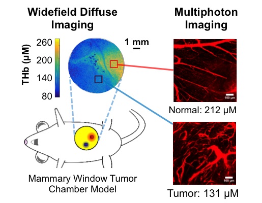
Multiscale imaging of mammary tumors. Spatial frequency domain imaging (SFDI) is used to measure widefield concentrations of hemoglobin, and multiphoton microcopy is used to quantify the morphology of tumor microvasculature. This combination of imaging modalities simultaneously provides micro-scale and widefield (clinically translatable) indications of chemotherapy response and resistance.
Publications:
M Applegate, Darren Roblyer, "High-speed Spatial Frequency Domain Imaging (SFDI) with temporally modulated light," Journal of Biomedical Optics, 22(7), 076019 (2017)
A. Torjesen, R Istfan, Darren Roblyer, "Ultrafast wavelength multiplexed broad bandwidth digital diffuse optical spectroscopy for in vivo extraction of tissue optical properties," Journal of Biomedical Optics, 22(3), 036009 (2017)
Link:
Last updated Wednesday, March 12, 2025