


Posted October 7, 2014
Kevin Cheung, M.D., Johns Hopkins University
 Ninety percent of breast cancers that become metastatic result in death. Invasion of tumor cells from the primary tumor to surrounding tissue is a fundamental step in metastasis. A clearer understanding of the cellular and molecular factors that underlie invasion could lead to breakthroughs in preventing invasion and, consequently, metastasis. The difficulty of monitoring tumor progression in vivo, however, poses a major challenge to elucidating the changes cancer cells undergo to execute invasion. Supported by an FY11 Postdoctoral Fellowship Award and co-mentored by Drs. Andrew Ewald and Sara Sukumar, Principal Investigator Dr. Kevin Cheung developed an assay to identify the most invasive cells within a primary tumor and then the unique molecular markers that could be exploited to target these cells and curb their invasive and metastatic potential.
Dr. Cheung and colleagues developed a three-dimensional (3D) organoid assay that models the architecture of breast tissue in greater detail than the more common 2D cultures. The assay utilizes "tumor organoids" - collections of cohesive tumor cells that preserve much of the cellular heterogeneity found in primary tumors - cultured within 3D collagen I gels, which models the tissue microenvironment surrounding invasive breast cancers. Using organoids generated from a breast cancer mouse model, the team observed that tumor organoids extended multicellular strands of cancer cells into the collagen I. Because the cells leading these invasive strands were highly protrusive and migratory, they were termed "invasive leader cells."
Molecular profiling of the leader cells showed enhanced expression of several basal epithelial markers including the transcription factor p63 and the intermediate filament protein cytokeratin-14 (K14). Tumor organoids triggered p63 and K14 expression before initiation of collective, multicellular invasion, indicating that these genes are components of a dynamically regulated gene program. Remarkably, while only a minority of cells within tumors expressed these genes, knockdown of basal epithelial gene expression was sufficient to block collective invasion in 3D cultures and in vivo.
Dr. Cheung and colleagues observed K14+ leader cells across major breast cancer subtypes, leading them to suggest that a shared molecular program for collective invasion may underlie the most common breast cancers. These data show that the heterogeneous interactions between tumor cell subpopulations are critical to collective invasion and that targeting the key drivers of invasion can significantly disrupt metastatic progression.
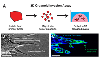
A. Tumor organoids are isolated from fresh primary breast tumors through a combination of mechanical disruption and enzymatic digestion. When tumor organoids are embedded in 3D collagen I matrix, they become invasive. B. Tumor organoids extend multiple invasive strands into the collagen matrix led by protrusive "invasive leader cells". C. Invasive leaders cells are molecularly distinct from the bulk tumor cells and express markers of basal differentiation, such as keratin-14 (K14). Scale bar is 50 microns.
Publications:
Cheung KJ, Gabrielson E, Werb Z, and Ewald AJ. 2013. Collective invasion in breast cancer requires a conserved basal epithelial program. Cell 155(7):1639-1651.
Nguyen-Ngoc KV, Cheung KJ, Brenot A, Shamir ER, Gray RS, Hines WC, Yaswen P, Werb Z, and Ewald AJ. 2012. ECM microenvironment regulates collective migration and local dissemination in normal and malignant mammary epithelium. Proc Natl Acad Sci U S A 109(39):E2595-604.