


Posted March 19, 2015
Sheng-Kwei Song, Ph.D., Washington University in St. Louis

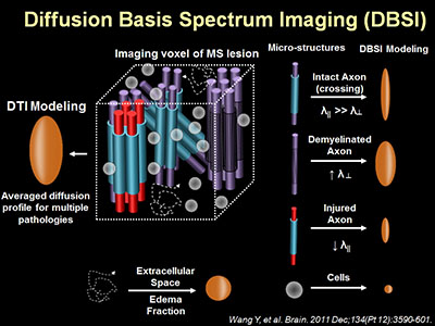
Multiple sclerosis (MS) is a common chronic inflammatory disease of the central nervous system (CNS), affecting about 0.1% of the population. It destroys myelin sheaths surrounding nerve fibers and in some cases leads to axonal loss. Although magnetic resonance imaging (MRI) has revolutionized the diagnosis of MS, it is currently unable to reveal the underlying complexities of the disease including inflammation, demyelination, and axonal loss. To overcome these limitations, Dr. Sheng-Kwei Song, with funding from a fiscal year 2011 Multiple Sclerosis Research Program Idea Award, is developing an advanced MRI technology called "diffusion basis spectrum imaging" (DBSI). The technique models tissue water diffusion characteristics in and around nerve axons and has the potential to identify different types of nerve lesions commonly observed in patients with MS.
To test this newly developed MRI technology, Dr. Song is imaging the brains of mice with experimentally induced nerve injuries that recapitulate MS lesions. The first model uses a neurotoxicant, cuprizone, to reversibly induce demyelination of nerve fibers with minimal axonal injury. Dr. Song demonstrated that the DBSI can detect increases in the space surrounding nerve axons that are associated with demyelinated nerve fibers. He also showed that these spaces decrease when the nerve fibers heal and become re-myelinated.
The second mouse model, autoimmune encephalomyelitis (EAE), more closely resembles the autoimmune nature of MS. The model results in an inflammatory demyelinating disease in the optic nerve (optic neuritis). He demonstrated that the DBSI technique can detect axonal injury and demyelination and quantify the extent of cellular inflammation and an increased accumulation of fluid (edema) in the optic nerve. This is important as edema and inflammatory cells obscure the ability of current imaging techniques to detect actual damage that occurs. The new technique enhances the use of imaging to determine the actual condition of nerves without the interference of other cellular response and thereby delivers a clearer picture of nerve health for diagnosis and enhances the ability to measure the effectiveness of potential therapies.
Dr. Song's short-term goal is to use this advanced MRI imaging technique to monitor the effectiveness of therapeutic drugs for MS in animal models, but the work also furthers his long-term goal of translating DBSI technology to noninvasively evaluate the underlying complexities of nerve damage seen in MS patients and of accelerating development of effective therapeutics for this disease.
Publications:
Wang X, Cusick MF, Wang Y, et al. 2014. Diffusion basis spectrum imaging detects and distinguishes coexisting subclinical inflammation, demyelination and axonal injury in experimental autoimmune encephalomyelitis mice. NMR Biomed 27(7):843-852.
Lin TH, Spees WM, Chiang CW, et al. 2014. Diffusion fMRI detects white-matter dysfunction in mice with acute optic neuritis. Neurobiol Dis 67:1-8.
Chiang CW, Wang Y, Sun P, et al. 2014. Quantifying white matter tract diffusion parameters in the presence of increased extra-fiber cellularity and vasogenic edema. Neuroimage 101:310-319.
Link: