


Posted March 6, 2014
Pedram Mohseni, Ph.D., Case Western Reserve University, Cleveland, Ohio
Randolph J. Nudo, Ph.D., University of Kansas Medical Center Research Institute, Kansas City, Kansas
 Loss of functionality associated with brain injury is not only caused by damage to critical brain regions but also by disruption of normal communications between undamaged areas. Dr. Pedram Mohseni at Case Western Reserve University and Dr. Randolph J. Nudo at the University of Kansas Medical Center Research Institute have teamed up to attempt to develop an artificial brain prosthesis that reestablishes communications between regions whose links are cut by brain injury. The investigators have successfully used their system to connect brain regions in rats and will soon attempt to perform the same sort of remediation in other animal models.
The rat has a grasping type of paw, and the researchers focused on disrupting and then restoring the communication links between sensory-motor regions in the brain that are required for the rat to perform a skilled reaching and grasping task. Rats were trained to reach through a narrow slot to retrieve a food pellet. In order for a rat to complete this task, there must be communication between the sensory brain region (dubbed S1) and the motor brain regions (dubbed CFA and RFA). Signals from S1 are processed by the CFA. The CFA and RFA together send signals through the spinal cord to move the limb and paw. Damage to the CFA region causes permanent loss of skilled grasping performance despite remaining connections between the RFA and spinal cord. This is presumably because the RFA lacks the connections with S1. (S1 = primary somatosensory cortex, CFA = caudal forelimb area, RFA = rostral forelimb area)
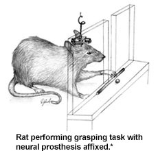 The researchers developed a miniaturized electronic processor that relays signals directly from the RFA area to the S1 area (skipping the damaged CFA) in hopes of restoring the sensory-motor communication links, thus restoring the grasping function when the CFA is injured. In these experiments, the CFA is precisely disrupted by a calibrated impactor, and electrodes are implanted into the S1 and RFA. The neuroprosthetic microdevice is connected to these electrodes and affixed to the top of the rat's skull. The battery-powered microdevice includes a wireless link that allows researchers to record signals, adjust operating parameters, and switch the device on and off to observe the difference in limb function when the device is in use versus turned off. The prosthesis uses a controller that distinguishes impulses in the RFA due to noise or artifacts from those signals that would normally be associated with volitional limb movement. When the prosthesis detects a movement signal in the RFA, a pulse is sent to S1 with the same delay that would normally have been associated with processing and passage through the CFA, and each pulse that is passed is followed by a short quiet period. Researchers call this activity-dependent or spike-triggered mode of stimulation "closed-loop" operation.
CFA-injured rats fitted with a closed-loop prosthesis regained near-normal levels of grasping skill by 15 days following injury, while CFA-injured rats with no prosthesis exhibited substantial loss of ability and no improvement over time. Notably, rats fitted with the prosthesis exhibited significantly better performance as early as day 3 following CFA injury (the first day of testing after injury).
Since repeated electrical stimulation of the brain is known to improve function after injury, the researchers sought to determine whether the observed return of grasping ability was simply due to the repeated stimulation. To do this the researchers fitted some rats with prostheses that operated in a random (or "open-loop") mode. These prostheses did not process signals from the RFA but simply sent pulses to S1 at random intervals that overall mimicked the average rate of stimuli delivered with the closed-loop system. These rats exhibited significant return of grasping performance but did not exhibit early return of function and ultimately only performed about half as well as the rats fitted with the closed-loop prostheses. This study demonstrates that closed-loop neuroprostheses can be used to improve communication in specific pathways in the injured brain, providing a possible new modality for treatment.
*Figure from Guggenmos DJ, Azin M, Barbay S, et al. 2013. Restoration of function after brain damage using a neural prosthesis. Proc Nat Acad Sci U S A 110(52):21177-21182.
Links: