 An official website of the United States government
 ) or https:// means you've safely connected to the .mil website. Share sensitive information only on official, secure websites.
Ovarian Cancer
Falloposcope: A Minimally Invasive Tool for Early Ovarian Cancer Detection


Posted September 21, 2023
Dr. Jennifer Barton, University of Arizona
 Dr. Jennifer Barton
Ovarian cancer is known as the deadliest female reproductive malignancy, as few symptoms show in the early stages of the disease.1 According to the Surveillance, Epidemiology, and End Results (SEER) Program of the National Cancer Institute, ovarian cancer rates are highest in women
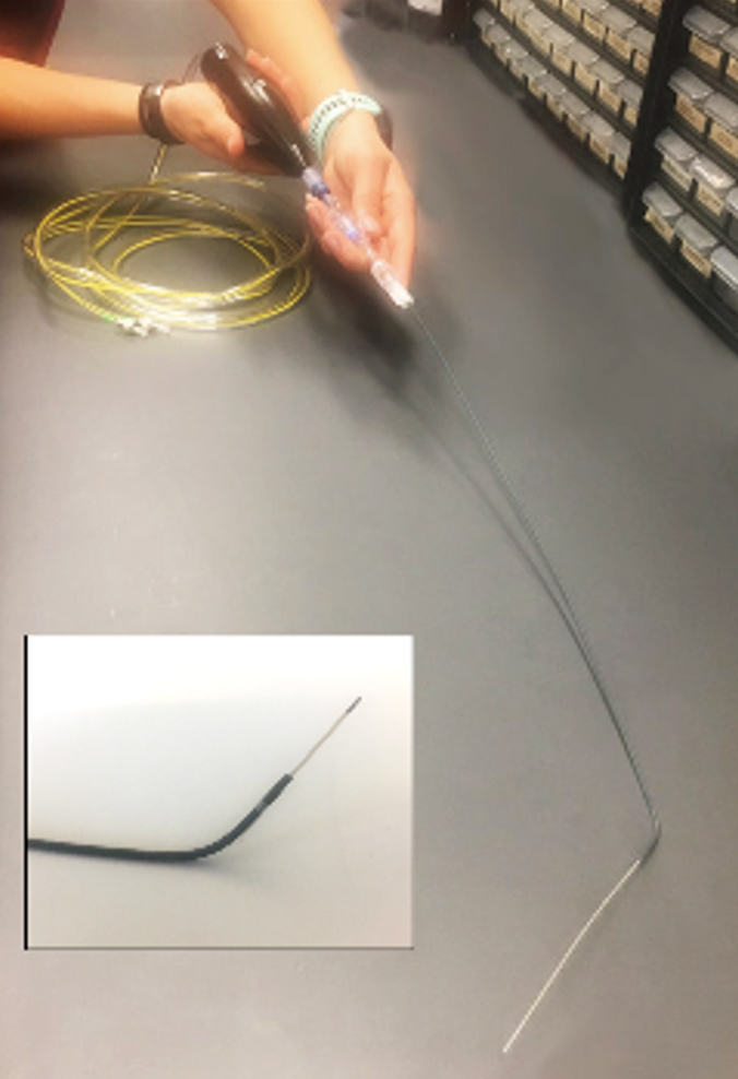 Figure 1. A researcher holds a minimally invasive falloposcope developed by Dr. Jennifer Barton, University of Arizona in Tucson, Arizona, though a FY17 Clinical Development Award from the Ovarian Cancer Research Program. Due to the small diameter of the scope, the imaging system causes no damage to the fallopian tubes.
Further, studies suggest that the deadliest ovarian cancer phenotypes originate in the fallopian tube, where a precursor lesion may dwell for years prior to metastasis to the ovary.3 Often, the disease is not treated until after spreading throughout the pelvis and abdomen, adding to the challenges for treatment or a cure.3 This serious condition for patients underscores the necessity for alternate imaging modalities and screening practices for the early detection of ovarian cancer.
Dr. Barton and her team designed a minimally invasive endoscope with advanced imaging techniques, including optical coherence tomography and multispectral fluorescence imaging, to scan the fallopian tubes for early signs of ovarian cancer and called the device as a falloposcope (Figure 1). Their novel imaging system can accurately detect small lesions in the fallopian tubes. Multiple procedural and equipment refinements made the imaging system quicker, compact, and more robust for optimal patient imaging. Although the COVID-19 pandemic initially delayed patient recruitment, Dr. Barton's team consented an initial cohort of 20 research individuals undergoing removal of their fallopian tubes for non-cancer reasons, ranging in age from
Dr. Barton's novel imaging system will enable the detection of pre-invasive cells, which will increase early intervention before cancerous cells disseminate from the fallopian tubes to the ovaries. Early detection of these precancerous cells will decrease the number of high-risk women undergoing prophylactic removal of their ovaries to prevent the disease. Since the ovaries provide health benefits even in post-menopausal women, the removal of these organs can increase mortality in women under 45 years old.2 This new imaging technology will ultimately improve the outcome for patients.
References:
1American Cancer Society. (2023). Key Statistics for Ovarian Cancer.
2SEER Stat Fact Sheets: Ovarian Cancer. (n.d.) National Cancer Institute, National Institute of Health.
3Bergsten, T. M., Burdette, J. E., & Dean, M. (2020). Fallopian tube initiation of high
Grade serous ovarian cancer and ovarian metastasis: Mechanisms and therapeutic
implications. Cancer letters, 476, 152:160. Retrieved from: https://doi.org/10.1016/
j.canlet.2020.02.017.
Links:
Last updated Monday, June 29, 2026